 Movie
Movie Controller

[English] 日本語
 Yorodumi
- PDB-8ab6: Complex III2 from Yarrowia lipolytica, combined datasets, consens... -
+ Open data

- Basic information
| Entry | Database: PDB / ID: 8ab6 | ||||||
|---|---|---|---|---|---|---|---|
| Title | Complex III2 from Yarrowia lipolytica, combined datasets, consensus refinement | ||||||
 Components Components |
| ||||||
 Keywords Keywords | MEMBRANE PROTEIN / oxidoreductase / electron transport chain | ||||||
| Function / homology |  Function and homology information Function and homology informationmitochondrial processing peptidase complex / mitochondrial processing peptidase / matrix side of mitochondrial inner membrane / : / mitochondrial respiratory chain complex III assembly / respiratory chain complex III / quinol-cytochrome-c reductase / quinol-cytochrome-c reductase activity / mitochondrial electron transport, ubiquinol to cytochrome c / nuclear periphery ...mitochondrial processing peptidase complex / mitochondrial processing peptidase / matrix side of mitochondrial inner membrane / : / mitochondrial respiratory chain complex III assembly / respiratory chain complex III / quinol-cytochrome-c reductase / quinol-cytochrome-c reductase activity / mitochondrial electron transport, ubiquinol to cytochrome c / nuclear periphery / metalloendopeptidase activity / mitochondrial intermembrane space / 2 iron, 2 sulfur cluster binding / oxidoreductase activity / mitochondrial inner membrane / heme binding / mitochondrion / proteolysis / membrane / metal ion binding Similarity search - Function | ||||||
| Biological species |  Yarrowia lipolytica (yeast) Yarrowia lipolytica (yeast) | ||||||
| Method | ELECTRON MICROSCOPY / single particle reconstruction / cryo EM / Resolution: 2 Å | ||||||
 Authors Authors | Wieferig, J.P. / Kuhlbrandt, W. | ||||||
| Funding support |  Germany, 1items Germany, 1items
| ||||||
 Citation Citation | Journal: IUCrJ / Year: 2023 Title: Analysis of the conformational heterogeneity of the Rieske iron-sulfur protein in complex III by cryo-EM. Authors: Jan Philip Wieferig / Werner Kühlbrandt / Abstract: Movement of the Rieske domain of the iron-sulfur protein is essential for intramolecular electron transfer within complex III (CIII) of the respiratory chain as it bridges a gap in the cofactor chain ...Movement of the Rieske domain of the iron-sulfur protein is essential for intramolecular electron transfer within complex III (CIII) of the respiratory chain as it bridges a gap in the cofactor chain towards the electron acceptor cytochrome c. We present cryo-EM structures of CIII from Yarrowia lipolytica at resolutions up to 2.0 Å under different conditions, with different redox states of the cofactors of the high-potential chain. All possible permutations of three primary positions were observed, indicating that the two halves of the dimeric complex act independently. Addition of the substrate analogue decylubiquinone to CIII with a reduced high-potential chain increased the occupancy of the Q site. The extent of Rieske domain interactions through hydrogen bonds to the cytochrome b and cytochrome c subunits varied depending on the redox state and substrate. In the absence of quinols, the reduced Rieske domain interacted more closely with cytochrome b and cytochrome c than in the oxidized state. Upon addition of the inhibitor antimycin A, the heterogeneity of the cd-helix and ef-loop increased, which may be indicative of a long-range effect on the Rieske domain. | ||||||
| History |
|
- Structure visualization
| Structure viewer | Molecule: MolmilJmol/JSmol |
|---|
- Downloads & links
-Download
| PDBx/mmCIF format | 8ab6.cif.gz | 843.4 KB | Display | PDBx/mmCIF format |
|---|---|---|---|---|
| PDB format | pdb8ab6.ent.gz | 664.5 KB | Display | PDB format |
| PDBx/mmJSON format | 8ab6.json.gz | Tree view | PDBx/mmJSON format | |
| Others |  Other downloads Other downloads |
-Validation report
| Arichive directory | https://data.pdbj.org/pub/pdb/validation_reports/ab/8ab6ftp://data.pdbj.org/pub/pdb/validation_reports/ab/8ab6 | HTTPS FTP |
|---|
-Related structure data
| Related structure data |  15312MC  8ab7C 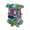 8ab8C  8ab9C  8abaC  8abbC  8abeC  8abfC  8abgC  8abhC  8abiC  8abjC 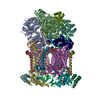 8abkC  8ablC  8abmC  8ac3C  8ac4C  8ac5C M: map data used to model this data C: citing same article ( |
|---|---|
| Similar structure data |


-Links
 PDBj
PDBj


- Assembly
| Deposited unit | 
| |||||||||||||||||||||||||||||||||||||||||||||||||||||||||||||||||||||||||||||||||||||||||||||||||||||||||||||||||||||||||||||||||||||||||||||||||||||||||||||||||||||||||||||||||||||||||||||||||||||||||
|---|---|---|---|---|---|---|---|---|---|---|---|---|---|---|---|---|---|---|---|---|---|---|---|---|---|---|---|---|---|---|---|---|---|---|---|---|---|---|---|---|---|---|---|---|---|---|---|---|---|---|---|---|---|---|---|---|---|---|---|---|---|---|---|---|---|---|---|---|---|---|---|---|---|---|---|---|---|---|---|---|---|---|---|---|---|---|---|---|---|---|---|---|---|---|---|---|---|---|---|---|---|---|---|---|---|---|---|---|---|---|---|---|---|---|---|---|---|---|---|---|---|---|---|---|---|---|---|---|---|---|---|---|---|---|---|---|---|---|---|---|---|---|---|---|---|---|---|---|---|---|---|---|---|---|---|---|---|---|---|---|---|---|---|---|---|---|---|---|---|---|---|---|---|---|---|---|---|---|---|---|---|---|---|---|---|---|---|---|---|---|---|---|---|---|---|---|---|---|---|---|---|---|
| 1 |
| |||||||||||||||||||||||||||||||||||||||||||||||||||||||||||||||||||||||||||||||||||||||||||||||||||||||||||||||||||||||||||||||||||||||||||||||||||||||||||||||||||||||||||||||||||||||||||||||||||||||||
| Noncrystallographic symmetry (NCS) | NCS domain:
NCS domain segments: Component-ID: 1 / Refine code: 1
|