 Movie
Movie Controller


+ Open data

- Basic information
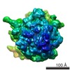
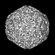
| Entry | Database: EMDB / ID: EMD-1466 | |||||||||
|---|---|---|---|---|---|---|---|---|---|---|
| Title | Human Parvovirus B19 (DNA-containing wildtype) | |||||||||
 Map data Map data | Human Parvovirus B19 (iB19, DNA-containing wildtype virions) | |||||||||
 Sample Sample |
| |||||||||
 Keywords Keywords | icosahedral virus / infectious / B19 | |||||||||
| Biological species |  Human parvovirus B19 Human parvovirus B19 | |||||||||
| Method | single particle reconstruction / cryo EM / Resolution: 7.5 Å | |||||||||
 Authors Authors | Kaufmann B / Chipman PR / Modrow S / Rossmann MG | |||||||||
 Citation Citation | Journal: J Virol / Year: 2008 Title: Visualization of the externalized VP2 N termini of infectious human parvovirus B19. Authors: Bärbel Kaufmann / Paul R Chipman / Victor A Kostyuchenko / Susanne Modrow / Michael G Rossmann /  Abstract: The structures of infectious human parvovirus B19 and empty wild-type particles were determined by cryoelectron microscopy (cryoEM) to 7.5-A and 11.3-A resolution, respectively, assuming icosahedral ...The structures of infectious human parvovirus B19 and empty wild-type particles were determined by cryoelectron microscopy (cryoEM) to 7.5-A and 11.3-A resolution, respectively, assuming icosahedral symmetry. Both of these, DNA filled and empty, wild-type particles contain a few copies of the minor capsid protein VP1. Comparison of wild-type B19 with the crystal structure and cryoEM reconstruction of recombinant B19 particles consisting of only the major capsid protein VP2 showed structural differences in the vicinity of the icosahedral fivefold axes. Although the unique N-terminal region of VP1 could not be visualized in the icosahedrally averaged maps, the N terminus of VP2 was shown to be exposed on the viral surface adjacent to the fivefold beta-cylinder. The conserved glycine-rich region is positioned between two neighboring, fivefold-symmetrically related VP subunits and not in the fivefold channel as observed for other parvoviruses. | |||||||||
| History |
|
- Structure visualization
| Movie |
 Movie viewer Movie viewer |
|---|---|
| Structure viewer | EM map: SurfViewMolmilJmol/JSmol |
| Supplemental images |

- Downloads & links
-EMDB archive
| Map data | emd_1466.map.gz | 2.5 MB | EMDB map data format | |
|---|---|---|---|---|
| Header (meta data) | emd-1466-v30.xmlemd-1466.xml | 10.3 KB 10.3 KB | Display Display | EMDB header |
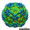
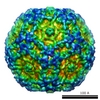
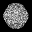
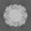
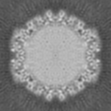
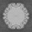
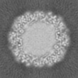
| Images | 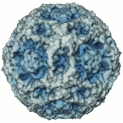 1466.gif 1466.gif | 114.5 KB | ||
| Archive directory |  http://ftp.pdbj.org/pub/emdb/structures/EMD-1466ftp://ftp.pdbj.org/pub/emdb/structures/EMD-1466 http://ftp.pdbj.org/pub/emdb/structures/EMD-1466ftp://ftp.pdbj.org/pub/emdb/structures/EMD-1466 | HTTPS FTP |
-Validation report
| Summary document | emd_1466_validation.pdf.gz | 239.4 KB | Display | EMDB validaton report |
|---|---|---|---|---|
| Full document | emd_1466_full_validation.pdf.gz | 238.5 KB | Display | |
| Data in XML | emd_1466_validation.xml.gz | 5.6 KB | Display | |
| Arichive directory | https://ftp.pdbj.org/pub/emdb/validation_reports/EMD-1466ftp://ftp.pdbj.org/pub/emdb/validation_reports/EMD-1466 | HTTPS FTP |
-Related structure data

-Links
| EMDB pages | EMDB (EBI/PDBe) / EMDataResource |
|---|
-Map
| File | Download / File: emd_1466.map.gz / Format: CCP4 / Size: 5.1 MB / Type: IMAGE STORED AS FLOATING POINT NUMBER (4 BYTES) | ||||||||||||||||||||||||||||||||||||||||||||||||||||||||||||||||||||
|---|---|---|---|---|---|---|---|---|---|---|---|---|---|---|---|---|---|---|---|---|---|---|---|---|---|---|---|---|---|---|---|---|---|---|---|---|---|---|---|---|---|---|---|---|---|---|---|---|---|---|---|---|---|---|---|---|---|---|---|---|---|---|---|---|---|---|---|---|---|
| Annotation | Human Parvovirus B19 (iB19, DNA-containing wildtype virions) | ||||||||||||||||||||||||||||||||||||||||||||||||||||||||||||||||||||
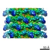
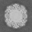
| Projections & slices | Image control
Images are generated by Spider. | ||||||||||||||||||||||||||||||||||||||||||||||||||||||||||||||||||||
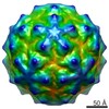
| Voxel size | X=Y=Z: 2.80054 Å | ||||||||||||||||||||||||||||||||||||||||||||||||||||||||||||||||||||
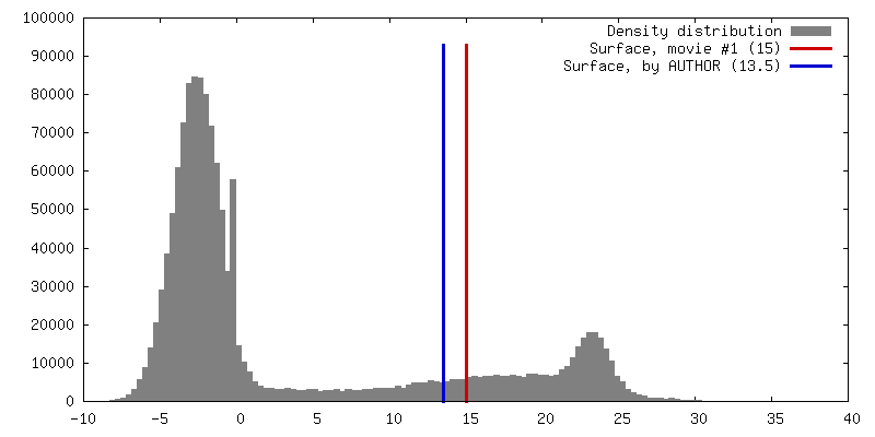
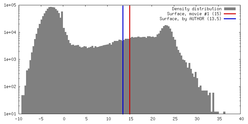
| Density |
| ||||||||||||||||||||||||||||||||||||||||||||||||||||||||||||||||||||
| Symmetry | Space group: 1 | ||||||||||||||||||||||||||||||||||||||||||||||||||||||||||||||||||||
| Details | EMDB XML:
CCP4 map header:
| ||||||||||||||||||||||||||||||||||||||||||||||||||||||||||||||||||||
 Z (Sec.)
 X (Row.)
 Y (Col.)


-Supplemental data
- Sample components
-Entire : Human Parvovirus B19 (DNA-containing wildtype particle)
| Entire | Name: Human Parvovirus B19 (DNA-containing wildtype particle) |
|---|---|
| Components |
|
-Supramolecule #1000: Human Parvovirus B19 (DNA-containing wildtype particle)
| Supramolecule | Name: Human Parvovirus B19 (DNA-containing wildtype particle) type: sample / ID: 1000 Details: icosahedral particle, DNA-containing virion, non-enveloped protein shell Oligomeric state: 60 VP subunits form icosahedral shell encapsidating 5.6kb ssDNA genome Number unique components: 1 |
|---|---|
| Molecular weight | Theoretical: 5.4 MDa |
-Supramolecule #1: Human parvovirus B19
| Supramolecule | Name: Human parvovirus B19 / type: virus / ID: 1 / Name.synonym: Human Parvovirus B19 / NCBI-ID: 10798 / Sci species name: Human parvovirus B19 / Virus type: VIRION / Virus isolate: SPECIES / Virus enveloped: No / Virus empty: No / Syn species name: Human Parvovirus B19 |
|---|---|
| Host (natural) | Organism:  Homo sapiens (human) / synonym: VERTEBRATES Homo sapiens (human) / synonym: VERTEBRATES |
| Molecular weight | Theoretical: 5.4 MDa |
| Virus shell | Shell ID: 1 / Diameter: 260 Å / T number (triangulation number): 1 |
-Experimental details
-Structure determination
| Method | cryo EM |
|---|---|
 Processing Processing | single particle reconstruction |
| Aggregation state | particle |
-Sample preparation
| Concentration | 1 mg/mL |
|---|---|
| Buffer | pH: 7.5 / Details: 25mM Tris-HCl |
| Grid | Details: 400 mesh copper |
| Vitrification | Cryogen name: ETHANE / Instrument: HOMEMADE PLUNGER Details: Vitrification instrument: guillotine-style plunge freezing device Method: A small vial of ethane is placed inside a larger liquid nitrogen reservoir. The grid holding a few microliters of the sample is held in place at the bottom of a plunger by the means of fine ...Method: A small vial of ethane is placed inside a larger liquid nitrogen reservoir. The grid holding a few microliters of the sample is held in place at the bottom of a plunger by the means of fine tweezers. Once the ethane in the vial is completely frozen, it needs to be slightly melted. When the liquid ethane is ready, a piece of filter paper is then pressed against the sample to blot of excess buffer, sufficient to leave a thin layer on the grid. After a predetermined time, the filter paper is removed, and the plunger is allowed to drop into the liquid ethane. Once the grid enters the liquid ethane, the sample is rapidly frozen, and the grid is transferred under liquid nitrogen to a storage box immersed liquid nitrogen for later use in the microscope. |
- Electron microscopy
| Microscope | FEI/PHILIPS CM300FEG/T |
|---|---|
| Temperature | Average: 98 K |
| Alignment procedure | Legacy - Astigmatism: live FFT at 200K |
| Details | low dose |
| Image recording | Category: FILM / Film or detector model: KODAK SO-163 FILM / Digitization - Scanner: ZEISS SCAI / Digitization - Sampling interval: 14 µm / Number real images: 50 / Average electron dose: 22 e/Å2 / Bits/pixel: 8 |
| Tilt angle min | 0 |
| Tilt angle max | 0 |
| Electron beam | Acceleration voltage: 300 kV / Electron source:  FIELD EMISSION GUN FIELD EMISSION GUN |
| Electron optics | Calibrated magnification: 47000 / Illumination mode: FLOOD BEAM / Imaging mode: BRIGHT FIELD / Cs: 2.0 mm / Nominal defocus max: 3.66 µm / Nominal defocus min: 1.25 µm / Nominal magnification: 45000 |
| Sample stage | Specimen holder: Eucentric / Specimen holder model: GATAN LIQUID NITROGEN |
-Image processing
| Details | manual particle selection |
|---|---|
| CTF correction | Details: each particle |
| Final reconstruction | Applied symmetry - Point group: I (icosahedral) / Algorithm: OTHER / Resolution.type: BY AUTHOR / Resolution: 7.5 Å / Resolution method: FSC 0.5 CUT-OFF / Software - Name: EMPFT, POR, P3DR Details: final map includes data to 7.15 Ang resolution (fsc 0.3 cut-off), magnification of final map standardized to a map calculated from B19 VP2 VLP model coordinates (PDB accession no 1S58) ...Details: final map includes data to 7.15 Ang resolution (fsc 0.3 cut-off), magnification of final map standardized to a map calculated from B19 VP2 VLP model coordinates (PDB accession no 1S58) resulting in final pixel separation of 2.8005Ang Number images used: 8854 |
