 Movie
Movie Controller


+ Open data

- Basic information
| Entry | Database: EMDB / ID: EMD-2244 | |||||||||
|---|---|---|---|---|---|---|---|---|---|---|
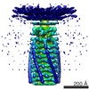
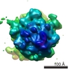
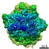
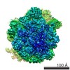
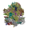
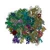
| Title | Cryo-electron microscopy of phirsl1 jumbo phage | |||||||||
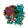
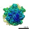
 Map data Map data | Reconstruction of the helical trunk of phirsl1 tail. A Bfactor of -538 has been applied to the reconstruction as well as a soft-edged mask. | |||||||||
 Sample Sample |
| |||||||||
 Keywords Keywords | Jumbo bacteriophage / phiRSL1 / Ralstonia solanacearum / cryo-electron microscopy / icosahedral capsid / helical tail / T6SS | |||||||||
| Function / homology | Tail sheath protein Function and homology information Function and homology information | |||||||||
| Biological species |  Ralstonia phage RSL1 (virus) Ralstonia phage RSL1 (virus) | |||||||||
| Method | helical reconstruction / cryo EM / negative staining / Resolution: 9.6 Å | |||||||||
 Authors Authors | Effantin G / Hamasaki R / Kawasaki T / Bacia M / Moriscot C / Weissenhorn W / Yamada T / Schoehn G | |||||||||
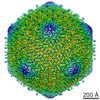
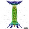
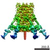
 Citation Citation | Journal: Structure / Year: 2013 Title: Cryo-electron microscopy three-dimensional structure of the jumbo phage ΦRSL1 infecting the phytopathogen Ralstonia solanacearum. Authors: Grégory Effantin / Ryosuke Hamasaki / Takeru Kawasaki / Maria Bacia / Christine Moriscot / Winfried Weissenhorn / Takashi Yamada / Guy Schoehn /  Abstract: ϕRSL1 jumbo phage belongs to a new class of viruses within the Myoviridae family. Here, we report its three-dimensional structure determined by electron cryo microscopy. The icosahedral capsid, the ...ϕRSL1 jumbo phage belongs to a new class of viruses within the Myoviridae family. Here, we report its three-dimensional structure determined by electron cryo microscopy. The icosahedral capsid, the tail helical portion, and the complete tail appendage were reconstructed separately to resolutions of 9 Å, 9 Å, and 28 Å, respectively. The head is rather complex and formed by at least five different proteins, whereas the major capsid proteins resemble those from HK97, despite low sequence conservation. The helical tail structure demonstrates its close relationship to T4 sheath proteins and provides evidence for an evolutionary link of the inner tail tube to the bacterial type VI secretion apparatus. Long fibers extend from the collar region, and their length is consistent with reaching the host cell surface upon tail contraction. Our structural analyses indicate that ϕRSL1 is an unusual member of the Myoviridae that employs conserved protein machines related to different phages and bacteria. | |||||||||
| History |
|
- Structure visualization
| Movie |
Movie viewer |
|---|---|
| Structure viewer | EM map: SurfViewMolmilJmol/JSmol |
| Supplemental images |

- Downloads & links
-EMDB archive
| Map data | emd_2244.map.gz | 1.8 MB | EMDB map data format | |
|---|---|---|---|---|
| Header (meta data) | emd-2244-v30.xmlemd-2244.xml | 10.4 KB 10.4 KB | Display Display | EMDB header |
| Images | 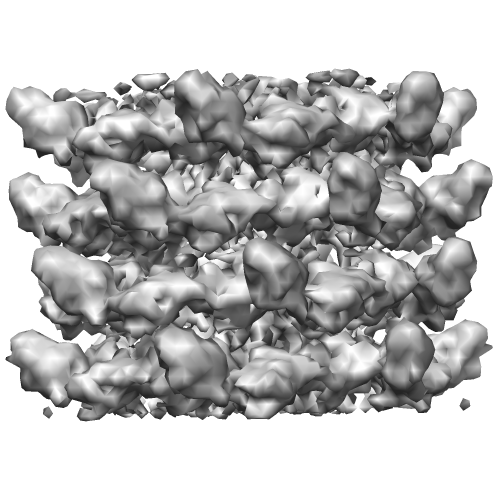 EMD-2244.png EMD-2244.png | 142.3 KB | ||
| Archive directory |  http://ftp.pdbj.org/pub/emdb/structures/EMD-2244ftp://ftp.pdbj.org/pub/emdb/structures/EMD-2244 http://ftp.pdbj.org/pub/emdb/structures/EMD-2244ftp://ftp.pdbj.org/pub/emdb/structures/EMD-2244 | HTTPS FTP |
-Related structure data

-Links
| EMDB pages | EMDB (EBI/PDBe) / EMDataResource |
|---|
-Map
| File | Download / File: emd_2244.map.gz / Format: CCP4 / Size: 6.4 MB / Type: IMAGE STORED AS FLOATING POINT NUMBER (4 BYTES) | ||||||||||||||||||||||||||||||||||||||||||||||||||||||||||||||||||||
|---|---|---|---|---|---|---|---|---|---|---|---|---|---|---|---|---|---|---|---|---|---|---|---|---|---|---|---|---|---|---|---|---|---|---|---|---|---|---|---|---|---|---|---|---|---|---|---|---|---|---|---|---|---|---|---|---|---|---|---|---|---|---|---|---|---|---|---|---|---|
| Annotation | Reconstruction of the helical trunk of phirsl1 tail. A Bfactor of -538 has been applied to the reconstruction as well as a soft-edged mask. | ||||||||||||||||||||||||||||||||||||||||||||||||||||||||||||||||||||
| Projections & slices | Image control
Images are generated by Spider. | ||||||||||||||||||||||||||||||||||||||||||||||||||||||||||||||||||||
| Voxel size | X=Y=Z: 2.26 Å | ||||||||||||||||||||||||||||||||||||||||||||||||||||||||||||||||||||
| Density |
| ||||||||||||||||||||||||||||||||||||||||||||||||||||||||||||||||||||
| Symmetry | Space group: 1 | ||||||||||||||||||||||||||||||||||||||||||||||||||||||||||||||||||||
| Details | EMDB XML:
CCP4 map header:
| ||||||||||||||||||||||||||||||||||||||||||||||||||||||||||||||||||||
 Z (Sec.)
 Y (Row.)
 X (Col.)


-Supplemental data
- Sample components
-Entire : phirsl1 helical portion of the tail
| Entire | Name: phirsl1 helical portion of the tail |
|---|---|
| Components |
|
-Supramolecule #1000: phirsl1 helical portion of the tail
| Supramolecule | Name: phirsl1 helical portion of the tail / type: sample / ID: 1000 / Number unique components: 2 |
|---|
-Macromolecule #1: Tail tube protein
| Macromolecule | Name: Tail tube protein / type: protein_or_peptide / ID: 1 / Details: UNP B2ZY00 / Recombinant expression: No / Database: NCBI |
|---|---|
| Source (natural) | Organism: Ralstonia phage RSL1 (virus) |
| Molecular weight | Theoretical: 18 KDa |
-Macromolecule #2: Tail sheath protein
| Macromolecule | Name: Tail sheath protein / type: protein_or_peptide / ID: 2 / Details: UNP B2ZXZ8 / Recombinant expression: No / Database: NCBI |
|---|---|
| Source (natural) | Organism: Ralstonia phage RSL1 (virus) |
| Molecular weight | Theoretical: 68 KDa |
| Sequence | InterPro: Tail sheath protein |
-Experimental details
-Structure determination
| Method | negative staining, cryo EM |
|---|---|
 Processing Processing | helical reconstruction |
| Aggregation state | filament |
-Sample preparation
| Buffer | pH: 7.5 / Details: 50 mM Tris-HCl [pH 7.5], 100 mM NaCl, 10 mM MgSO4 |
|---|---|
| Staining | Type: NEGATIVE Details: Four microliters of the bacteriophage sample (~0.1 mg/ml) were loaded between the mica-carbon interface. The sample was stained using 2% ammonium molybdate pH 7.5 and air-dried. |
| Vitrification | Cryogen name: ETHANE / Chamber humidity: 100 % / Instrument: FEI VITROBOT MARK IV |
- Electron microscopy
| Microscope | FEI POLARA 300 |
|---|---|
| Alignment procedure | Legacy - Astigmatism: Objective lens astigmatism was corrected at 59000 |
| Date | Feb 25, 2011 |
| Image recording | Category: FILM / Film or detector model: KODAK SO-163 FILM / Digitization - Scanner: ZEISS SCAI / Digitization - Sampling interval: 7 µm / Number real images: 52 / Average electron dose: 20 e/Å2 / Bits/pixel: 8 |
| Electron beam | Acceleration voltage: 300 kV / Electron source:  FIELD EMISSION GUN FIELD EMISSION GUN |
| Electron optics | Illumination mode: SPOT SCAN / Imaging mode: BRIGHT FIELD / Cs: 2.3 mm / Nominal defocus max: 5.295 µm / Nominal defocus min: 1.355 µm / Nominal magnification: 31000 |
| Sample stage | Specimen holder model: GATAN LIQUID NITROGEN |
| Experimental equipment |  Model: Tecnai Polara / Image courtesy: FEI Company |
-Image processing
| Details | Projection matching was done with SPIDER. IHRSR was used for helical parameters search and application. |
|---|---|
| Final reconstruction | Applied symmetry - Helical parameters - Δz: 37.9 Å Applied symmetry - Helical parameters - Δ&Phi: 22.04 ° Applied symmetry - Helical parameters - Axial symmetry: C6 (6 fold cyclic) Algorithm: OTHER / Resolution.type: BY AUTHOR / Resolution: 9.6 Å / Resolution method: FSC 0.5 CUT-OFF / Software - Name: SPIDER, IHRSR |
| CTF correction | Details: each particle |
