ムービー
ムービー コントローラー


+ データを開く

- 基本情報
基本情報
| 登録情報 | データベース: EMDB / ID: EMD-13441 | ||||||||||||
|---|---|---|---|---|---|---|---|---|---|---|---|---|---|
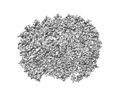
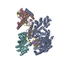
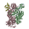
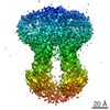
| タイトル | Cryo-EM structure of the Rhodobacter sphaeroides RC-LH1-PufXY monomer complex at 2.5 A | ||||||||||||
 マップデータ マップデータ | |||||||||||||
 試料 試料 |
| ||||||||||||
 キーワード キーワード | Photosynthetic bacteria / light harvesting complex / Cryo-EM / RC-LH1 / RC-LH1-PufX / RC-LH1-PufX-Y / PHOTOSYNTHESIS | ||||||||||||
| 機能・相同性 |  機能・相同性情報 機能・相同性情報organelle inner membrane / plasma membrane-derived chromatophore membrane / plasma membrane light-harvesting complex / bacteriochlorophyll binding / : / photosynthetic electron transport in photosystem II / photosynthesis, light reaction / photosynthesis / metal ion binding / membrane / plasma membrane 類似検索 - 分子機能 | ||||||||||||
| 生物種 |  Cereibacter sphaeroides 2.4.1 (バクテリア) / Rhodobacter sphaeroides (strain ATCC 17023 / DSM 158 / JCM 6121 / NBRC 12203 / NCIMB 8253 / ATH 2.4.1.) (バクテリア) / Rhodobacter sphaeroides (strain ATCC 17023 / 2.4.1 / NCIB 8253 / DSM 158) (バクテリア) Cereibacter sphaeroides 2.4.1 (バクテリア) / Rhodobacter sphaeroides (strain ATCC 17023 / DSM 158 / JCM 6121 / NBRC 12203 / NCIMB 8253 / ATH 2.4.1.) (バクテリア) / Rhodobacter sphaeroides (strain ATCC 17023 / 2.4.1 / NCIB 8253 / DSM 158) (バクテリア) | ||||||||||||
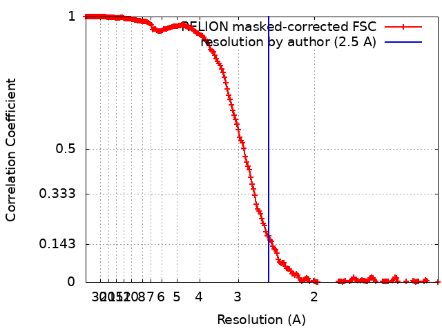
| 手法 | 単粒子再構成法 / クライオ電子顕微鏡法 / 解像度: 2.5 Å | ||||||||||||
 データ登録者 データ登録者 | Qian P / Hunter CN | ||||||||||||
| 資金援助 |  英国, 3件 英国, 3件
| ||||||||||||
 引用 引用 | ジャーナル: Biochem J / 年: 2021 タイトル: Cryo-EM structure of the monomeric Rhodobacter sphaeroides RC-LH1 core complex at 2.5 Å. 著者: Pu Qian / David J K Swainsbury / Tristan I Croll / Jack H Salisbury / Elizabeth C Martin / Philip J Jackson / Andrew Hitchcock / Pablo Castro-Hartmann / Kasim Sader / C Neil Hunter /  要旨: Reaction centre light-harvesting 1 (RC-LH1) complexes are the essential components of bacterial photosynthesis. The membrane-intrinsic LH1 complex absorbs light and the energy migrates to an enclosed ...Reaction centre light-harvesting 1 (RC-LH1) complexes are the essential components of bacterial photosynthesis. The membrane-intrinsic LH1 complex absorbs light and the energy migrates to an enclosed RC where a succession of electron and proton transfers conserves the energy as a quinol, which is exported to the cytochrome bc1 complex. In some RC-LH1 variants quinols can diffuse through small pores in a fully circular, 16-subunit LH1 ring, while in others missing LH1 subunits create a gap for quinol export. We used cryogenic electron microscopy to obtain a 2.5 Å resolution structure of one such RC-LH1, a monomeric complex from Rhodobacter sphaeroides. The structure shows that the RC is partly enclosed by a 14-subunit LH1 ring in which each αβ heterodimer binds two bacteriochlorophylls and, unusually for currently reported complexes, two carotenoids rather than one. Although the extra carotenoids confer an advantage in terms of photoprotection and light harvesting, they could impede passage of quinones through small, transient pores in the LH1 ring, necessitating a mechanism to create a dedicated quinone channel. The structure shows that two transmembrane proteins play a part in stabilising an open ring structure; one of these components, the PufX polypeptide, is augmented by a hitherto undescribed protein subunit we designate as protein-Y, which lies against the transmembrane regions of the thirteenth and fourteenth LH1α polypeptides. Protein-Y prevents LH1 subunits 11-14 adjacent to the RC QB site from bending inwards towards the RC and, with PufX preventing complete encirclement of the RC, this pair of polypeptides ensures unhindered quinone diffusion. #1: ジャーナル: Acta Crystallogr., Sect. D: Biol. Crystallogr.年: 2018 タイトル: Real-space refinement in PHENIX for cryo-EM and crystallography 著者: qian P / Hunter CN #2: ジャーナル: To Be Publishedタイトル: Cryo-EM structure of the Rhodobacter sphaeroides RC-LH1-PufXY monomer complex at 2.5 A 著者: Qian P / Hunter CN | ||||||||||||
| 履歴 |
|
- 構造の表示
構造の表示
| ムービー |
ムービービューア |
|---|---|
| 構造ビューア | EMマップ: SurfViewMolmilJmol/JSmol |
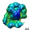
| 添付画像 |
- ダウンロードとリンク
-EMDBアーカイブ
| マップデータ | emd_13441.map.gz | 37.1 MB | EMDBマップデータ形式 | |
|---|---|---|---|---|
| ヘッダ (付随情報) | emd-13441-v30.xmlemd-13441.xml | 25.2 KB 25.2 KB | 表示 表示 | EMDBヘッダ |
| FSC (解像度算出) | emd_13441_fsc.xml | 18.2 KB | 表示 | FSCデータファイル |
| 画像 |  emd_13441.png emd_13441.png | 108.3 KB | ||
| Filedesc metadata | emd-13441.cif.gz | 7.1 KB | ||
| アーカイブディレクトリ |  http://ftp.pdbj.org/pub/emdb/structures/EMD-13441ftp://ftp.pdbj.org/pub/emdb/structures/EMD-13441 http://ftp.pdbj.org/pub/emdb/structures/EMD-13441ftp://ftp.pdbj.org/pub/emdb/structures/EMD-13441 | HTTPS FTP |
-関連構造データ


-リンク
| EMDBのページ | EMDB (EBI/PDBe) / EMDataResource |
|---|---|
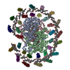
| 「今月の分子」の関連する項目 |


-マップ
| ファイル | ダウンロード / ファイル: emd_13441.map.gz / 形式: CCP4 / 大きさ: 512 MB / タイプ: IMAGE STORED AS FLOATING POINT NUMBER (4 BYTES) | ||||||||||||||||||||||||||||||||||||||||||||||||||||||||||||||||||||
|---|---|---|---|---|---|---|---|---|---|---|---|---|---|---|---|---|---|---|---|---|---|---|---|---|---|---|---|---|---|---|---|---|---|---|---|---|---|---|---|---|---|---|---|---|---|---|---|---|---|---|---|---|---|---|---|---|---|---|---|---|---|---|---|---|---|---|---|---|---|
| 投影像・断面図 | 画像のコントロール
画像は Spider により作成 | ||||||||||||||||||||||||||||||||||||||||||||||||||||||||||||||||||||
| ボクセルのサイズ | X=Y=Z: 0.65 Å | ||||||||||||||||||||||||||||||||||||||||||||||||||||||||||||||||||||
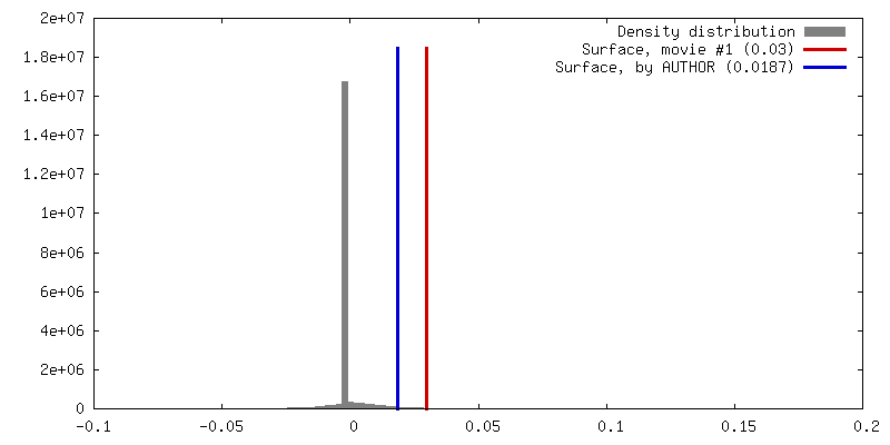
| 密度 |
| ||||||||||||||||||||||||||||||||||||||||||||||||||||||||||||||||||||
| 対称性 | 空間群: 1 | ||||||||||||||||||||||||||||||||||||||||||||||||||||||||||||||||||||
| 詳細 | EMDB XML:
CCP4マップ ヘッダ情報:
| ||||||||||||||||||||||||||||||||||||||||||||||||||||||||||||||||||||
 Z (Sec.)
 Y (Row.)
 X (Col.)


-添付データ
- 試料の構成要素
+全体 : Light harvesting complex
+超分子 #1: Light harvesting complex
+分子 #1: Light-harvesting protein B-875 alpha chain
+分子 #2: Light-harvesting protein B-875 beta chain
+分子 #3: Reaction center protein H chain
+分子 #4: Reaction center protein L chain
+分子 #5: Reaction center protein M chain
+分子 #6: RC-Y
+分子 #7: Intrinsic membrane protein PufX
+分子 #8: BACTERIOCHLOROPHYLL A

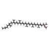
+分子 #9: SPHEROIDENE
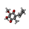
+分子 #10: DODECYL-BETA-D-MALTOSIDE

+分子 #11: 1,2-Distearoyl-sn-glycerophosphoethanolamine

+分子 #12: BACTERIOPHEOPHYTIN A

+分子 #13: UBIQUINONE-10

+分子 #14: UBIQUINONE-1
+分子 #15: (2R,5R,11R,14R)-5,8,11-trihydroxy-5,11-dioxido-17-oxo-2,14-bis(te...

+分子 #16: FE (III) ION
+分子 #17: water

-実験情報
-構造解析
| 手法 | クライオ電子顕微鏡法 |
|---|---|
 解析 解析 | 単粒子再構成法 |
| 試料の集合状態 | particle |
-試料調製
| 濃度 | 4.0 mg/mL |
|---|---|
| 緩衝液 | pH: 7.8 / 構成要素 - 濃度: 20.0 mMol / 構成要素 - 式: HEPES / 詳細: 20 mM HEPES, pH 7.8 , 0.03% beta-DDM |
| グリッド | モデル: Quantifoil R1.2/1.3 / 材質: COPPER / メッシュ: 300 / 前処理 - 雰囲気: OTHER |
| 凍結 | 凍結剤: ETHANE / チャンバー内湿度: 100 % / チャンバー内温度: 277 K / 装置: FEI VITROBOT MARK III / 詳細: QF R1.2/1.3 grid coated graphene oxide. |
| 詳細 | in 0.03% beta-DDM detergent |
- 電子顕微鏡法
| 顕微鏡 | FEI TITAN KRIOS |
|---|---|
| 撮影 | フィルム・検出器のモデル: FEI FALCON IV (4k x 4k) 撮影したグリッド数: 1 / 実像数: 3180 / 平均露光時間: 12.21 sec. / 平均電子線量: 44.94 e/Å2 |
| 電子線 | 加速電圧: 300 kV / 電子線源:  FIELD EMISSION GUN FIELD EMISSION GUN |
| 電子光学系 | C2レンズ絞り径: 50.0 µm / 照射モード: FLOOD BEAM / 撮影モード: BRIGHT FIELD / Cs: 2.7 mm / 最大 デフォーカス(公称値): 2.2 µm / 最小 デフォーカス(公称値): 0.8 µm / 倍率(公称値): 120000 |
| 試料ステージ | 試料ホルダーモデル: FEI TITAN KRIOS AUTOGRID HOLDER ホルダー冷却材: NITROGEN |
| 実験機器 |  モデル: Titan Krios / 画像提供: FEI Company |