+ データを開く
- 基本情報
基本情報
| 登録情報 | データベース: EMDB / ID: EMD-1321 | |||||||||
|---|---|---|---|---|---|---|---|---|---|---|
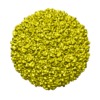
| タイトル | Quasi-atomic model of bacteriophage t7 procapsid shell: insights into the structure and evolution of a basic fold. | |||||||||
 マップデータ マップデータ | phage T7 prohead icosahedral map | |||||||||
 試料 試料 |
| |||||||||
| 機能・相同性 | Capsid Gp10A/Gp10B / : / Major capsid protein / viral capsid / viral translational frameshifting / identical protein binding / Major capsid protein 機能・相同性情報 機能・相同性情報 | |||||||||
| 生物種 |   Enterobacteria phage T7 (ファージ) Enterobacteria phage T7 (ファージ) | |||||||||
| 手法 | 単粒子再構成法 / クライオ電子顕微鏡法 / 解像度: 10.9 Å | |||||||||
 データ登録者 データ登録者 | Agirrezabala X / Velazquez-Muriel J / Gomez-Puertas P / Scheres S / Carazo JM / Carrascosa JL | |||||||||
 引用 引用 |  ジャーナル: Structure / 年: 2007 ジャーナル: Structure / 年: 2007タイトル: Quasi-atomic model of bacteriophage t7 procapsid shell: insights into the structure and evolution of a basic fold. 著者: Xabier Agirrezabala / Javier A Velázquez-Muriel / Paulino Gómez-Puertas / Sjors H W Scheres / José M Carazo / José L Carrascosa /  要旨: The existence of similar folds among major structural subunits of viral capsids has shown unexpected evolutionary relationships suggesting common origins irrespective of the capsids' host life domain. ...The existence of similar folds among major structural subunits of viral capsids has shown unexpected evolutionary relationships suggesting common origins irrespective of the capsids' host life domain. Tailed bacteriophages are emerging as one such family, and we have studied the possible existence of the HK97-like fold in bacteriophage T7. The procapsid structure at approximately 10 A resolution was used to obtain a quasi-atomic model by fitting a homology model of the T7 capsid protein gp10 that was based on the atomic structure of the HK97 capsid protein. A number of fold similarities, such as the fitting of domains A and P into the L-shaped procapsid subunit, are evident between both viral systems. A different feature is related to the presence of the amino-terminal domain of gp10 found at the inner surface of the capsid that might play an important role in the interaction of capsid and scaffolding proteins. | |||||||||
| 履歴 |
|
- 構造の表示
構造の表示
| ムービー |
 ムービービューア ムービービューア |
|---|---|
| 構造ビューア | EMマップ:  SurfView SurfView Molmil Molmil Jmol/JSmol Jmol/JSmol |
| 添付画像 |
- ダウンロードとリンク
-EMDBアーカイブ
| マップデータ |  emd_1321.map.gz emd_1321.map.gz | 6.8 MB |  EMDBマップデータ形式 EMDBマップデータ形式 | |
|---|---|---|---|---|
| ヘッダ (付随情報) |  emd-1321-v30.xml emd-1321-v30.xml emd-1321.xml emd-1321.xml | 8.4 KB 8.4 KB | 表示 表示 |  EMDBヘッダ EMDBヘッダ |
| 画像 |  1321.gif 1321.gif | 85.7 KB | ||
| アーカイブディレクトリ |  http://ftp.pdbj.org/pub/emdb/structures/EMD-1321 http://ftp.pdbj.org/pub/emdb/structures/EMD-1321 ftp://ftp.pdbj.org/pub/emdb/structures/EMD-1321 ftp://ftp.pdbj.org/pub/emdb/structures/EMD-1321 | HTTPS FTP |
-検証レポート
| 文書・要旨 |  emd_1321_validation.pdf.gz emd_1321_validation.pdf.gz | 281.2 KB | 表示 |  EMDB検証レポート EMDB検証レポート |
|---|---|---|---|---|
| 文書・詳細版 |  emd_1321_full_validation.pdf.gz emd_1321_full_validation.pdf.gz | 280.4 KB | 表示 | |
| XML形式データ |  emd_1321_validation.xml.gz emd_1321_validation.xml.gz | 6.3 KB | 表示 | |
| アーカイブディレクトリ |  https://ftp.pdbj.org/pub/emdb/validation_reports/EMD-1321 https://ftp.pdbj.org/pub/emdb/validation_reports/EMD-1321 ftp://ftp.pdbj.org/pub/emdb/validation_reports/EMD-1321 ftp://ftp.pdbj.org/pub/emdb/validation_reports/EMD-1321 | HTTPS FTP |
-関連構造データ
- リンク
リンク
| EMDBのページ |  EMDB (EBI/PDBe) / EMDB (EBI/PDBe) /  EMDataResource EMDataResource |
|---|
- マップ
マップ
| ファイル |  ダウンロード / ファイル: emd_1321.map.gz / 形式: CCP4 / 大きさ: 65.5 MB / タイプ: IMAGE STORED AS FLOATING POINT NUMBER (4 BYTES) ダウンロード / ファイル: emd_1321.map.gz / 形式: CCP4 / 大きさ: 65.5 MB / タイプ: IMAGE STORED AS FLOATING POINT NUMBER (4 BYTES) | ||||||||||||||||||||||||||||||||||||||||||||||||||||||||||||||||||||
|---|---|---|---|---|---|---|---|---|---|---|---|---|---|---|---|---|---|---|---|---|---|---|---|---|---|---|---|---|---|---|---|---|---|---|---|---|---|---|---|---|---|---|---|---|---|---|---|---|---|---|---|---|---|---|---|---|---|---|---|---|---|---|---|---|---|---|---|---|---|
| 注釈 | phage T7 prohead icosahedral map | ||||||||||||||||||||||||||||||||||||||||||||||||||||||||||||||||||||
| 投影像・断面図 | 画像のコントロール
画像は Spider により作成 | ||||||||||||||||||||||||||||||||||||||||||||||||||||||||||||||||||||
| ボクセルのサイズ | X=Y=Z: 2.72 Å | ||||||||||||||||||||||||||||||||||||||||||||||||||||||||||||||||||||
| 密度 |
| ||||||||||||||||||||||||||||||||||||||||||||||||||||||||||||||||||||
| 対称性 | 空間群: 1 | ||||||||||||||||||||||||||||||||||||||||||||||||||||||||||||||||||||
| 詳細 | EMDB XML:
CCP4マップ ヘッダ情報:
| ||||||||||||||||||||||||||||||||||||||||||||||||||||||||||||||||||||
-添付データ
- 試料の構成要素
-全体 : phage T7 prohead
| 全体 | 名称: phage T7 prohead |
|---|---|
| 要素 |
|
-超分子 #1000: phage T7 prohead
| 超分子 | 名称: phage T7 prohead / タイプ: sample / ID: 1000 / Number unique components: 1 |
|---|
-分子 #1: gp10A
| 分子 | 名称: gp10A / タイプ: protein_or_peptide / ID: 1 / Name.synonym: major capsid protein / コピー数: 420 / 集合状態: icosahedral / 組換発現: Yes |
|---|---|
| 由来(天然) | 生物種:   Enterobacteria phage T7 (ファージ) / 別称: phage T7 Enterobacteria phage T7 (ファージ) / 別称: phage T7 |
| 分子量 | 理論値: 37 MDa |
| 組換発現 | 生物種:  |
-実験情報
-構造解析
| 手法 | クライオ電子顕微鏡法 |
|---|---|
 解析 解析 | 単粒子再構成法 |
| 試料の集合状態 | particle |
- 試料調製
試料調製
| 緩衝液 | pH: 7.7 / 詳細: 50mM Tris-HCl pH:7.7 10mM MgCl2 100mM NaCl |
|---|---|
| グリッド | 詳細: Quantifoil grids 2/2 |
| 凍結 | 凍結剤: ETHANE |
- 電子顕微鏡法
| 顕微鏡 | FEI TECNAI 20 |
|---|---|
| アライメント法 | Legacy - 非点収差: 100k |
| 撮影 | カテゴリ: FILM / フィルム・検出器のモデル: KODAK SO-163 FILM / デジタル化 - スキャナー: ZEISS SCAI / デジタル化 - サンプリング間隔: 7 µm / 平均電子線量: 10 e/Å2 / ビット/ピクセル: 8 |
| Tilt angle min | 0 |
| Tilt angle max | 0 |
| 電子線 | 加速電圧: 200 kV / 電子線源:  FIELD EMISSION GUN FIELD EMISSION GUN |
| 電子光学系 | 倍率(補正後): 51600 / 照射モード: SPOT SCAN / 撮影モード: BRIGHT FIELD / Cs: 2.26 mm / 最大 デフォーカス(公称値): 3.0 µm / 最小 デフォーカス(公称値): 1.0 µm / 倍率(公称値): 50000 |
| 試料ステージ | 試料ホルダー: Side entry liquid nitrogen-cooled cryo specimen holder. GATAN. Eucentric 試料ホルダーモデル: GATAN LIQUID NITROGEN |
- 画像解析
画像解析
| CTF補正 | 詳細: Wiener filter, defocus groups |
|---|---|
| 最終 再構成 | 想定した対称性 - 点群: I (正20面体型対称) / アルゴリズム: OTHER / 解像度のタイプ: BY AUTHOR / 解像度: 10.9 Å / 解像度の算出法: FSC 0.5 CUT-OFF / ソフトウェア - 名称: spider / 使用した粒子像数: 4460 |
 ムービー
ムービー コントローラー


 Z (Sec.)
 Y (Row.)
 X (Col.)


