[English] 日本語
 Yorodumi
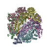
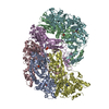
- EMDB-13148: Human mitochondrial Lon protease with substrate in the ATPase and... -
+ Open data
- Basic information
| Entry | Database: EMDB / ID: EMD-13148 | |||||||||
|---|---|---|---|---|---|---|---|---|---|---|
| Title | Human mitochondrial Lon protease with substrate in the ATPase and protease domains | |||||||||
 Map data Map data | Human mitochondrial LonP1 K898A, LocScale map | |||||||||
 Sample Sample |
| |||||||||
 Keywords Keywords | Protease / Mitochondria / AAA+ / HYDROLASE | |||||||||
| Function / homology |  Function and homology information Function and homology informationoxidation-dependent protein catabolic process / response to aluminum ion / PH domain binding / endopeptidase La / mitochondrial protein catabolic process / mitochondrial DNA metabolic process / G-quadruplex DNA binding / : / ATP-dependent peptidase activity / protein quality control for misfolded or incompletely synthesized proteins ...oxidation-dependent protein catabolic process / response to aluminum ion / PH domain binding / endopeptidase La / mitochondrial protein catabolic process / mitochondrial DNA metabolic process / G-quadruplex DNA binding / : / ATP-dependent peptidase activity / protein quality control for misfolded or incompletely synthesized proteins / mitochondrial nucleoid / insulin receptor substrate binding / Mitochondrial unfolded protein response (UPRmt) / chaperone-mediated protein complex assembly / response to hormone / DNA polymerase binding / negative regulation of insulin receptor signaling pathway / Mitochondrial protein degradation / : / mitochondrion organization / protein catabolic process / ADP binding / single-stranded DNA binding / cellular response to oxidative stress / sequence-specific DNA binding / response to hypoxia / single-stranded RNA binding / mitochondrial matrix / serine-type endopeptidase activity / ATP hydrolysis activity / mitochondrion / nucleoplasm / ATP binding / identical protein binding / membrane / cytosol Similarity search - Function | |||||||||
| Biological species |  Homo sapiens (human) Homo sapiens (human) | |||||||||
| Method | single particle reconstruction / cryo EM / Resolution: 2.75 Å | |||||||||
 Authors Authors | Valentin Gese G / Shahzad S | |||||||||
 Citation Citation |  Journal: To Be Published Journal: To Be PublishedTitle: A dual allosteric pathway drives human mitochondrial Lon Authors: Valentin Gese G / Shahzad S / Pardo-Hernandez C / Wramstedt A / Falkenberg M / Hallberg M | |||||||||
| History |
|
- Structure visualization
| Movie |
 Movie viewer Movie viewer |
|---|---|
| Structure viewer | EM map:  SurfView SurfView Molmil Molmil Jmol/JSmol Jmol/JSmol |
| Supplemental images |
- Downloads & links
-EMDB archive
| Map data |  emd_13148.map.gz emd_13148.map.gz | 405.3 MB |  EMDB map data format EMDB map data format | |
|---|---|---|---|---|
| Header (meta data) |  emd-13148-v30.xml emd-13148-v30.xml emd-13148.xml emd-13148.xml | 18.1 KB 18.1 KB | Display Display |  EMDB header EMDB header |
| FSC (resolution estimation) |  emd_13148_fsc.xml emd_13148_fsc.xml | 20.9 KB | Display |  FSC data file FSC data file |
| Images |  emd_13148.png emd_13148.png | 78.9 KB | ||
| Masks |  emd_13148_msk_1.map emd_13148_msk_1.map | 824 MB |  Mask map Mask map | |
| Filedesc metadata |  emd-13148.cif.gz emd-13148.cif.gz | 5.9 KB | ||
| Others |  emd_13148_additional_1.map.gz emd_13148_additional_1.map.gz emd_13148_half_map_1.map.gz emd_13148_half_map_1.map.gz emd_13148_half_map_2.map.gz emd_13148_half_map_2.map.gz | 410.2 MB 763.7 MB 763.7 MB | ||
| Archive directory |  http://ftp.pdbj.org/pub/emdb/structures/EMD-13148 http://ftp.pdbj.org/pub/emdb/structures/EMD-13148 ftp://ftp.pdbj.org/pub/emdb/structures/EMD-13148 ftp://ftp.pdbj.org/pub/emdb/structures/EMD-13148 | HTTPS FTP |
-Validation report
| Summary document |  emd_13148_validation.pdf.gz emd_13148_validation.pdf.gz | 1.1 MB | Display |  EMDB validaton report EMDB validaton report |
|---|---|---|---|---|
| Full document |  emd_13148_full_validation.pdf.gz emd_13148_full_validation.pdf.gz | 1.1 MB | Display | |
| Data in XML |  emd_13148_validation.xml.gz emd_13148_validation.xml.gz | 29.7 KB | Display | |
| Data in CIF |  emd_13148_validation.cif.gz emd_13148_validation.cif.gz | 38.9 KB | Display | |
| Arichive directory |  https://ftp.pdbj.org/pub/emdb/validation_reports/EMD-13148 https://ftp.pdbj.org/pub/emdb/validation_reports/EMD-13148 ftp://ftp.pdbj.org/pub/emdb/validation_reports/EMD-13148 ftp://ftp.pdbj.org/pub/emdb/validation_reports/EMD-13148 | HTTPS FTP |
-Related structure data
| Related structure data |  7p0mMC  7p09C  7p0bC M: atomic model generated by this map C: citing same article ( |
|---|---|
| Similar structure data |
- Links
Links
| EMDB pages |  EMDB (EBI/PDBe) / EMDB (EBI/PDBe) /  EMDataResource EMDataResource |
|---|---|
| Related items in Molecule of the Month |
- Map
Map
| File |  Download / File: emd_13148.map.gz / Format: CCP4 / Size: 824 MB / Type: IMAGE STORED AS FLOATING POINT NUMBER (4 BYTES) Download / File: emd_13148.map.gz / Format: CCP4 / Size: 824 MB / Type: IMAGE STORED AS FLOATING POINT NUMBER (4 BYTES) | ||||||||||||||||||||||||||||||||||||||||||||||||||||||||||||||||||||
|---|---|---|---|---|---|---|---|---|---|---|---|---|---|---|---|---|---|---|---|---|---|---|---|---|---|---|---|---|---|---|---|---|---|---|---|---|---|---|---|---|---|---|---|---|---|---|---|---|---|---|---|---|---|---|---|---|---|---|---|---|---|---|---|---|---|---|---|---|---|
| Annotation | Human mitochondrial LonP1 K898A, LocScale map | ||||||||||||||||||||||||||||||||||||||||||||||||||||||||||||||||||||
| Projections & slices | Image control
Images are generated by Spider. | ||||||||||||||||||||||||||||||||||||||||||||||||||||||||||||||||||||
| Voxel size | X=Y=Z: 0.654 Å | ||||||||||||||||||||||||||||||||||||||||||||||||||||||||||||||||||||
| Density |
| ||||||||||||||||||||||||||||||||||||||||||||||||||||||||||||||||||||
| Symmetry | Space group: 1 | ||||||||||||||||||||||||||||||||||||||||||||||||||||||||||||||||||||
| Details | EMDB XML:
CCP4 map header:
| ||||||||||||||||||||||||||||||||||||||||||||||||||||||||||||||||||||
-Supplemental data
-Mask #1
| File |  emd_13148_msk_1.map emd_13148_msk_1.map | ||||||||||||
|---|---|---|---|---|---|---|---|---|---|---|---|---|---|
| Projections & Slices |
| ||||||||||||
| Density Histograms |
-Additional map: Human mitochondrial LonP1 K898A
| File | emd_13148_additional_1.map | ||||||||||||
|---|---|---|---|---|---|---|---|---|---|---|---|---|---|
| Annotation | Human mitochondrial LonP1 K898A | ||||||||||||
| Projections & Slices |
| ||||||||||||
| Density Histograms |
-Half map: Human mitochondrial LonP1 K898A, half map B
| File | emd_13148_half_map_1.map | ||||||||||||
|---|---|---|---|---|---|---|---|---|---|---|---|---|---|
| Annotation | Human mitochondrial LonP1 K898A, half map B | ||||||||||||
| Projections & Slices |
| ||||||||||||
| Density Histograms |
-Half map: Human mitochondrial LonP1 K898A, half map A
| File | emd_13148_half_map_2.map | ||||||||||||
|---|---|---|---|---|---|---|---|---|---|---|---|---|---|
| Annotation | Human mitochondrial LonP1 K898A, half map A | ||||||||||||
| Projections & Slices |
| ||||||||||||
| Density Histograms |
- Sample components
-Entire : Human mitochondrial LonP1 K898A mutant
| Entire | Name: Human mitochondrial LonP1 K898A mutant |
|---|---|
| Components |
|
-Supramolecule #1: Human mitochondrial LonP1 K898A mutant
| Supramolecule | Name: Human mitochondrial LonP1 K898A mutant / type: complex / ID: 1 / Parent: 0 / Macromolecule list: #1-#2 |
|---|---|
| Source (natural) | Organism:  Homo sapiens (human) Homo sapiens (human) |
| Molecular weight | Theoretical: 591 KDa |
-Macromolecule #1: Lon protease homolog, mitochondrial
| Macromolecule | Name: Lon protease homolog, mitochondrial / type: protein_or_peptide / ID: 1 / Number of copies: 6 / Enantiomer: LEVO / EC number: endopeptidase La |
|---|---|
| Source (natural) | Organism:  Homo sapiens (human) Homo sapiens (human) |
| Molecular weight | Theoretical: 99.713266 KDa |
| Recombinant expression | Organism:  |
| Sequence | String: SMGFWEASSR GGGAFSGGED ASEGGAEEGA GGAGGSAGAG EGPVITALTP MTIPDVFPHL PLIAITRNPV FPRFIKIIEV KNKKLVELL RRKVRLAQPY VGVFLKRDDS NESDVVESLD EIYHTGTFAQ IHEMQDLGDK LRMIVMGHRR VHISRQLEVE P EEPEAENK ...String: SMGFWEASSR GGGAFSGGED ASEGGAEEGA GGAGGSAGAG EGPVITALTP MTIPDVFPHL PLIAITRNPV FPRFIKIIEV KNKKLVELL RRKVRLAQPY VGVFLKRDDS NESDVVESLD EIYHTGTFAQ IHEMQDLGDK LRMIVMGHRR VHISRQLEVE P EEPEAENK HKPRRKSKRG KKEAEDELSA RHPAELAMEP TPELPAEVLM VEVENVVHED FQVTEEVKAL TAEIVKTIRD II ALNPLYR ESVLQMMQAG QRVVDNPIYL SDMGAALTGA ESHELQDVLE ETNIPKRLYK ALSLLKKEFE LSKLQQRLGR EVE EKIKQT HRKYLLQEQL KIIKKELGLE KDDKDAIEEK FRERLKELVV PKHVMDVVDE ELSKLGLLDN HSSEFNVTRN YLDW LTSIP WGKYSNENLD LARAQAVLEE DHYGMEDVKK RILEFIAVSQ LRGSTQGKIL CFYGPPGVGK TSIARSIARA LNREY FRFS VGGMTDVAEI KGHRRTYVGA MPGKIIQCLK KTKTENPLIL IDEVDKIGRG YQGDPSSALL ELLDPEQNAN FLDHYL DVP VDLSKVLFIC TANVTDTIPE PLRDRMEMIN VSGYVAQEKL AIAERYLVPQ ARALCGLDES KAKLSSDVLT LLIKQYC RE SGVRNLQKQV EKVLRKSAYK IVSGEAESVE VTPENLQDFV GKPVFTVERM YDVTPPGVVM GLAWTAMGGS TLFVETSL R RPQDKDAKGD KDGSLEVTGQ LGEVMKESAR IAYTFARAFL MQHAPANDYL VTSHIHLHVP EGATPKDGPS AGCTIVTAL LSLAMGRPVR QNLAMTGEVS LTGKILPVGG IKEATIAAKR AGVTCIVLPA ENKKDFYDLA AFITEGLEVH FVEHYREIFD IAFPDEQAE ALAVER UniProtKB: Lon protease homolog, mitochondrial |
-Macromolecule #2: Unknown peptide from human mitochondrial transcription factor A (TFAM)
| Macromolecule | Name: Unknown peptide from human mitochondrial transcription factor A (TFAM) type: protein_or_peptide / ID: 2 / Number of copies: 7 / Enantiomer: LEVO |
|---|---|
| Source (natural) | Organism:  Homo sapiens (human) Homo sapiens (human) |
| Molecular weight | Theoretical: 954.168 Da |
| Recombinant expression | Organism:  |
| Sequence | String: (UNK)(UNK)(UNK)(UNK)(UNK)(UNK)(UNK)(UNK)(UNK)(UNK) (UNK) |
-Macromolecule #3: ADENOSINE-5'-TRIPHOSPHATE
| Macromolecule | Name: ADENOSINE-5'-TRIPHOSPHATE / type: ligand / ID: 3 / Number of copies: 4 / Formula: ATP |
|---|---|
| Molecular weight | Theoretical: 507.181 Da |
| Chemical component information |  ChemComp-ATP: |
-Macromolecule #4: MAGNESIUM ION
| Macromolecule | Name: MAGNESIUM ION / type: ligand / ID: 4 / Number of copies: 4 / Formula: MG |
|---|---|
| Molecular weight | Theoretical: 24.305 Da |
-Macromolecule #5: ADENOSINE-5'-DIPHOSPHATE
| Macromolecule | Name: ADENOSINE-5'-DIPHOSPHATE / type: ligand / ID: 5 / Number of copies: 2 / Formula: ADP |
|---|---|
| Molecular weight | Theoretical: 427.201 Da |
| Chemical component information |  ChemComp-ADP: |
-Experimental details
-Structure determination
| Method | cryo EM |
|---|---|
 Processing Processing | single particle reconstruction |
| Aggregation state | particle |
- Sample preparation
| Buffer | pH: 7.5 |
|---|---|
| Vitrification | Cryogen name: ETHANE |
- Electron microscopy
| Microscope | FEI TITAN KRIOS |
|---|---|
| Image recording | Film or detector model: GATAN K3 BIOQUANTUM (6k x 4k) / Average exposure time: 1.5 sec. / Average electron dose: 51.0 e/Å2 |
| Electron beam | Acceleration voltage: 300 kV / Electron source:  FIELD EMISSION GUN FIELD EMISSION GUN |
| Electron optics | Illumination mode: SPOT SCAN / Imaging mode: BRIGHT FIELD |
| Experimental equipment |  Model: Titan Krios / Image courtesy: FEI Company |
 Movie
Movie Controller


 Z (Sec.)
 Y (Row.)
 X (Col.)


