 ムービー
ムービー コントローラー


+ データを開く

- 基本情報
基本情報
| 登録情報 |  | ||||||||||||||||||
|---|---|---|---|---|---|---|---|---|---|---|---|---|---|---|---|---|---|---|---|
| タイトル | Cryo-EM structure of the 6 hpf zebrafish embryo 80S ribosome | ||||||||||||||||||
 マップデータ マップデータ | |||||||||||||||||||
 試料 試料 |
| ||||||||||||||||||
| 機能・相同性 |  機能・相同性情報 機能・相同性情報optokinetic behavior / L13a-mediated translational silencing of Ceruloplasmin expression / SRP-dependent cotranslational protein targeting to membrane / RMTs methylate histone arginines / TNFR1-induced NF-kappa-B signaling pathway / TNFR1-mediated ceramide production / : / : / Formation of a pool of free 40S subunits / Formation of the ternary complex, and subsequently, the 43S complex ...optokinetic behavior / L13a-mediated translational silencing of Ceruloplasmin expression / SRP-dependent cotranslational protein targeting to membrane / RMTs methylate histone arginines / TNFR1-induced NF-kappa-B signaling pathway / TNFR1-mediated ceramide production / : / : / Formation of a pool of free 40S subunits / Formation of the ternary complex, and subsequently, the 43S complex / Ribosomal scanning and start codon recognition / : / Protein methylation / Nonsense Mediated Decay (NMD) independent of the Exon Junction Complex (EJC) / Nonsense Mediated Decay (NMD) enhanced by the Exon Junction Complex (EJC) / mTORC1-mediated signalling / brain segmentation / Regulation of TNFR1 signaling / convergent extension involved in gastrulation / : / : / primitive hemopoiesis / embryonic retina morphogenesis in camera-type eye / anatomical structure development / hemoglobin biosynthetic process / mitochondrial cytochrome c oxidase assembly / chordate embryonic development / embryonic brain development / ribosomal subunit / exocrine pancreas development / positive regulation of gastrulation / positive regulation of translational fidelity / mitochondrial large ribosomal subunit / definitive hemopoiesis / pancreas development / regulation of establishment of cell polarity / cytoplasmic side of rough endoplasmic reticulum membrane / laminin receptor activity / negative regulation of Wnt signaling pathway / regulation of innate immune response / regulation of cell division / erythrocyte maturation / hemopoiesis / intrinsic apoptotic signaling pathway by p53 class mediator / endonucleolytic cleavage to generate mature 3'-end of SSU-rRNA from (SSU-rRNA, 5.8S rRNA, LSU-rRNA) / mitochondrial respiratory chain complex I assembly / hematopoietic stem cell differentiation / protein-RNA complex assembly / vasculogenesis / erythrocyte development / translation regulator activity / laminin binding / rough endoplasmic reticulum / endonucleolytic cleavage in ITS1 to separate SSU-rRNA from 5.8S rRNA and LSU-rRNA from tricistronic rRNA transcript (SSU-rRNA, 5.8S rRNA, LSU-rRNA) / class I DNA-(apurinic or apyrimidinic site) endonuclease activity / DNA-(apurinic or apyrimidinic site) lyase / rescue of stalled ribosome / maturation of LSU-rRNA from tricistronic rRNA transcript (SSU-rRNA, 5.8S rRNA, LSU-rRNA) / maturation of SSU-rRNA from tricistronic rRNA transcript (SSU-rRNA, 5.8S rRNA, LSU-rRNA) / erythrocyte differentiation / maturation of LSU-rRNA / ribosomal large subunit biogenesis / maturation of SSU-rRNA / small-subunit processome / positive regulation of apoptotic signaling pathway / protein kinase C binding / regulation of erythrocyte differentiation / brain development / ribosomal large subunit assembly / mRNA 5'-UTR binding / modification-dependent protein catabolic process / spindle / rRNA processing / large ribosomal subunit / protein tag activity / regulation of protein localization / ribosome binding / glucose homeostasis / regulation of translation / ribosomal small subunit biogenesis / ribosomal small subunit assembly / small ribosomal subunit / small ribosomal subunit rRNA binding / 5S rRNA binding / T cell differentiation in thymus / large ribosomal subunit rRNA binding / cytosolic small ribosomal subunit / angiogenesis / cytosolic large ribosomal subunit / mitochondrial inner membrane / cytoplasmic translation / rRNA binding / negative regulation of translation / ribosome / regulation of cell cycle / protein ubiquitination / structural constituent of ribosome / ribonucleoprotein complex / translation / positive regulation of protein phosphorylation 類似検索 - 分子機能 | ||||||||||||||||||
| 生物種 |  | ||||||||||||||||||
| 手法 | 単粒子再構成法 / クライオ電子顕微鏡法 / 解像度: 2.4 Å | ||||||||||||||||||
 データ登録者 データ登録者 | Leesch F / Lorenzo-Orts L / Grishkovskaya I / Kandolf S / Belacic K / Meinhart A / Haselbach D / Pauli A | ||||||||||||||||||
| 資金援助 |  オーストリア, オーストリア,  スイス, 5件 スイス, 5件
| ||||||||||||||||||
 引用 引用 | ジャーナル: Nature / 年: 2023 タイトル: A molecular network of conserved factors keeps ribosomes dormant in the egg. 著者: Friederike Leesch / Laura Lorenzo-Orts / Carina Pribitzer / Irina Grishkovskaya / Josef Roehsner / Anastasia Chugunova / Manuel Matzinger / Elisabeth Roitinger / Katarina Belačić / Susanne ...著者: Friederike Leesch / Laura Lorenzo-Orts / Carina Pribitzer / Irina Grishkovskaya / Josef Roehsner / Anastasia Chugunova / Manuel Matzinger / Elisabeth Roitinger / Katarina Belačić / Susanne Kandolf / Tzi-Yang Lin / Karl Mechtler / Anton Meinhart / David Haselbach / Andrea Pauli / 要旨: Ribosomes are produced in large quantities during oogenesis and are stored in the egg. However, the egg and early embryo are translationally repressed. Here, using mass spectrometry and cryo-electron ...Ribosomes are produced in large quantities during oogenesis and are stored in the egg. However, the egg and early embryo are translationally repressed. Here, using mass spectrometry and cryo-electron microscopy analyses of ribosomes isolated from zebrafish (Danio rerio) and Xenopus laevis eggs and embryos, we provide molecular evidence that ribosomes transition from a dormant state to an active state during the first hours of embryogenesis. Dormant ribosomes are associated with four conserved factors that form two modules, consisting of Habp4-eEF2 and death associated protein 1b (Dap1b) or Dap in complex with eIF5a. Both modules occupy functionally important sites and act together to stabilize ribosomes and repress translation. Dap1b (also known as Dapl1 in mammals) is a newly discovered translational inhibitor that stably inserts into the polypeptide exit tunnel. Addition of recombinant zebrafish Dap1b protein is sufficient to block translation and reconstitute the dormant egg ribosome state in a mammalian translation extract in vitro. Thus, a developmentally programmed, conserved ribosome state has a key role in ribosome storage and translational repression in the egg. | ||||||||||||||||||
| 履歴 |
|
- 構造の表示
構造の表示
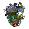
| 添付画像 |
|---|

- ダウンロードとリンク
-EMDBアーカイブ
| マップデータ | emd_13112.map.gz | 311.3 MB | EMDBマップデータ形式 | |
|---|---|---|---|---|
| ヘッダ (付随情報) | emd-13112-v30.xmlemd-13112.xml | 91.5 KB 91.5 KB | 表示 表示 | EMDBヘッダ |
| 画像 | 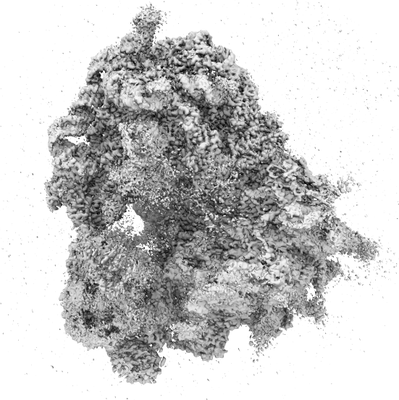 emd_13112.png emd_13112.png | 87.5 KB | ||
| アーカイブディレクトリ |  http://ftp.pdbj.org/pub/emdb/structures/EMD-13112ftp://ftp.pdbj.org/pub/emdb/structures/EMD-13112 http://ftp.pdbj.org/pub/emdb/structures/EMD-13112ftp://ftp.pdbj.org/pub/emdb/structures/EMD-13112 | HTTPS FTP |
-検証レポート
| 文書・要旨 | emd_13112_validation.pdf.gz | 432.1 KB | 表示 | EMDB検証レポート |
|---|---|---|---|---|
| 文書・詳細版 | emd_13112_full_validation.pdf.gz | 431.7 KB | 表示 | |
| XML形式データ | emd_13112_validation.xml.gz | 7.8 KB | 表示 | |
| CIF形式データ | emd_13112_validation.cif.gz | 9 KB | 表示 | |
| アーカイブディレクトリ | https://ftp.pdbj.org/pub/emdb/validation_reports/EMD-13112ftp://ftp.pdbj.org/pub/emdb/validation_reports/EMD-13112 | HTTPS FTP |
-関連構造データ


-リンク
| EMDBのページ | EMDB (EBI/PDBe) / EMDataResource |
|---|---|
| 「今月の分子」の関連する項目 |


-マップ
| ファイル | ダウンロード / ファイル: emd_13112.map.gz / 形式: CCP4 / 大きさ: 421.9 MB / タイプ: IMAGE STORED AS FLOATING POINT NUMBER (4 BYTES) | ||||||||||||||||||||
|---|---|---|---|---|---|---|---|---|---|---|---|---|---|---|---|---|---|---|---|---|---|
| ボクセルのサイズ | X=Y=Z: 1.06 Å | ||||||||||||||||||||
| 密度 |
| ||||||||||||||||||||
| 対称性 | 空間群: 1 | ||||||||||||||||||||
| 詳細 | EMDB XML:
|
-添付データ
- 試料の構成要素
+全体 : 80S ribosome from 6 hpf zebrafish embryos
+超分子 #1: 80S ribosome from 6 hpf zebrafish embryos
+分子 #1: 18S rRNA
+分子 #2: 28S rRNA
+分子 #3: 5S rRNA
+分子 #4: 5.8S rRNA
+分子 #5: 60S ribosomal protein L8
+分子 #6: 40S ribosomal protein SA
+分子 #7: Ribosomal protein L3
+分子 #8: 40S ribosomal protein S3a
+分子 #9: Ribosomal protein L4
+分子 #10: 40S ribosomal protein S2
+分子 #11: Ribosomal protein L5b
+分子 #12: DNA-(apurinic or apyrimidinic site) lyase
+分子 #13: 60S ribosomal protein L6
+分子 #14: 40S ribosomal protein S4, X isoform
+分子 #15: Ribosomal protein L7
+分子 #16: Ribosomal protein S5
+分子 #17: 60S ribosomal protein L7a
+分子 #18: 40S ribosomal protein S6
+分子 #19: 60S ribosomal protein L9
+分子 #20: 40S ribosomal protein S7
+分子 #21: 60S ribosomal protein L10
+分子 #22: 40S ribosomal protein S8
+分子 #23: 60S ribosomal protein L11
+分子 #24: 40S ribosomal protein S9
+分子 #25: Ribosomal protein S10
+分子 #26: 60S ribosomal protein L13
+分子 #27: 40S ribosomal protein S11
+分子 #28: 60S ribosomal protein L14
+分子 #29: Ribosomal protein L15
+分子 #30: 40S ribosomal protein S13
+分子 #31: 60S ribosomal protein L13a
+分子 #32: Ribosomal protein S14
+分子 #33: 60S ribosomal protein L17
+分子 #34: 40S ribosomal protein S15
+分子 #35: Ribosomal protein L18
+分子 #36: Ribosomal protein S16
+分子 #37: 60S ribosomal protein L19
+分子 #38: 40S ribosomal protein S17
+分子 #39: 60S ribosomal protein L18a
+分子 #40: 40S ribosomal protein S18
+分子 #41: 60S ribosomal protein L21
+分子 #42: 40S ribosomal protein S19
+分子 #43: Ribosomal protein L22
+分子 #44: 40S ribosomal protein S20
+分子 #45: 60S ribosomal protein L23
+分子 #46: 40S ribosomal protein S21
+分子 #47: 60S ribosomal protein L24
+分子 #48: 40S ribosomal protein S15a
+分子 #49: Ribosomal protein L23a
+分子 #50: 40S ribosomal protein S23
+分子 #51: ATPase H+ transporting V0 subunit e1
+分子 #52: 40S ribosomal protein S24
+分子 #53: 60S ribosomal protein L27
+分子 #54: 40S ribosomal protein S25
+分子 #55: 60S ribosomal protein L27a
+分子 #56: 40S ribosomal protein S26
+分子 #57: 60S ribosomal protein L29
+分子 #58: 40S ribosomal protein S27
+分子 #59: 60S ribosomal protein L30
+分子 #60: 40S ribosomal protein S28
+分子 #61: 60S ribosomal protein L31
+分子 #62: 40S ribosomal protein S29
+分子 #63: Ribosomal protein L32
+分子 #64: 40S ribosomal protein S30
+分子 #65: 60S ribosomal protein L35a
+分子 #66: 60S ribosomal protein L34
+分子 #67: Guanine nucleotide-binding protein subunit beta-2-like 1
+分子 #68: 60S ribosomal protein L35
+分子 #69: 60S ribosomal protein L36
+分子 #70: Ribosomal protein L37
+分子 #71: 60S ribosomal protein L38
+分子 #72: Ribosomal protein L39
+分子 #73: 60S ribosomal protein L40
+分子 #74: Rpl41
+分子 #75: 60S ribosomal protein L36a
+分子 #76: Zgc:171772
+分子 #77: 60S ribosomal protein L28
+分子 #78: MAGNESIUM ION
+分子 #79: ZINC ION
-実験情報
-構造解析
| 手法 | クライオ電子顕微鏡法 |
|---|---|
 解析 解析 | 単粒子再構成法 |
| 試料の集合状態 | particle |
-試料調製
| 緩衝液 | pH: 7.6 構成要素:
| ||||||||||||||||||
|---|---|---|---|---|---|---|---|---|---|---|---|---|---|---|---|---|---|---|---|
| グリッド | モデル: Quantifoil R3.5/1 / 材質: COPPER / メッシュ: 200 / 支持フィルム - 材質: CARBON / 支持フィルム - トポロジー: CONTINUOUS / 支持フィルム - Film thickness: 2.0 nm / 前処理 - タイプ: GLOW DISCHARGE / 前処理 - 時間: 60 sec. | ||||||||||||||||||
| 凍結 | 凍結剤: ETHANE / チャンバー内湿度: 70 % / 装置: LEICA EM GP |
- 電子顕微鏡法
| 顕微鏡 | FEI TITAN KRIOS |
|---|---|
| 撮影 | フィルム・検出器のモデル: FEI FALCON III (4k x 4k) 撮影したグリッド数: 1 / 実像数: 11860 / 平均電子線量: 48.3 e/Å2 |
| 電子線 | 加速電圧: 300 kV / 電子線源:  FIELD EMISSION GUN FIELD EMISSION GUN |
| 電子光学系 | 照射モード: FLOOD BEAM / 撮影モード: BRIGHT FIELD / Cs: 2.7 mm |
| 試料ステージ | 試料ホルダーモデル: FEI TITAN KRIOS AUTOGRID HOLDER ホルダー冷却材: NITROGEN |
| 実験機器 |  モデル: Titan Krios / 画像提供: FEI Company |
+画像解析
-原子モデル構築 1
| 精密化 | プロトコル: OTHER |
|---|---|
| 得られたモデル |  PDB-7oyb: |
