Movie
Movie Controller

[English] 日本語
 Yorodumi
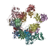
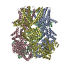
- EMDB-12638: Structure of the fungal plasma membrane proton pump Pma1 in its a... -
+ Open data

- Basic information
| Entry | Database: EMDB / ID: EMD-12638 | ||||||||||||||||||
|---|---|---|---|---|---|---|---|---|---|---|---|---|---|---|---|---|---|---|---|
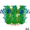
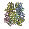
| Title | Structure of the fungal plasma membrane proton pump Pma1 in its auto-inhibited state - monomer unit | ||||||||||||||||||
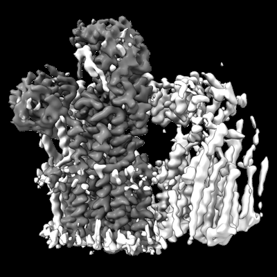
 Map data Map data | monomeric map derived from focused 3D classification and refinement of symmetry expanded particles | ||||||||||||||||||
 Sample Sample |
| ||||||||||||||||||
 Keywords Keywords | Membrane Protein / P-Type ATPase / proton-transporting ATPase / PROTON TRANSPORT | ||||||||||||||||||
| Function / homology |  Function and homology information Function and homology informationP-type H+-exporting transporter / proton export across plasma membrane / P-type proton-exporting transporter activity / ATP hydrolysis activity / ATP binding / plasma membrane Similarity search - Function | ||||||||||||||||||
| Biological species |  Neurospora crassa (fungus) Neurospora crassa (fungus) | ||||||||||||||||||
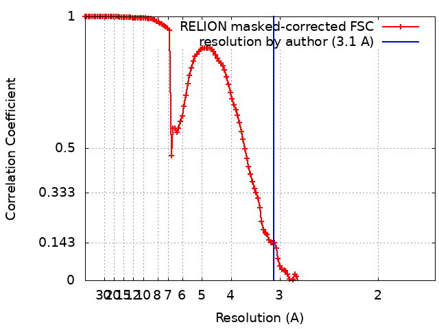
| Method | single particle reconstruction / cryo EM / Resolution: 3.1 Å | ||||||||||||||||||
 Authors Authors | Heit S / Geurts MMG | ||||||||||||||||||
| Funding support |  United Kingdom, 5 items United Kingdom, 5 items
| ||||||||||||||||||
 Citation Citation | Journal: Sci Adv / Year: 2021 Title: Structure of the hexameric fungal plasma membrane proton pump in its autoinhibited state. Authors: Sabine Heit / Maxwell M G Geurts / Bonnie J Murphy / Robin A Corey / Deryck J Mills / Werner Kühlbrandt / Maike Bublitz /  Abstract: The fungal plasma membrane H-ATPase Pma1 is a vital enzyme, generating a proton-motive force that drives the import of essential nutrients. Autoinhibited Pma1 hexamers in the plasma membrane of ...The fungal plasma membrane H-ATPase Pma1 is a vital enzyme, generating a proton-motive force that drives the import of essential nutrients. Autoinhibited Pma1 hexamers in the plasma membrane of starving fungi are activated by glucose signaling and subsequent phosphorylation of the autoinhibitory domain. As related P-type adenosine triphosphatases (ATPases) are not known to oligomerize, the physiological relevance of Pma1 hexamers remained unknown. We have determined the structure of hexameric Pma1 from by electron cryo-microscopy at 3.3-Å resolution, elucidating the molecular basis for hexamer formation and autoinhibition and providing a basis for structure-based drug development. Coarse-grained molecular dynamics simulations in a lipid bilayer suggest lipid-mediated contacts between monomers and a substantial protein-induced membrane deformation that could act as a proton-attracting funnel. | ||||||||||||||||||
| History |
|
- Structure visualization
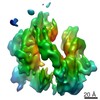
| Movie |
Movie viewer |
|---|---|
| Structure viewer | EM map: SurfViewMolmilJmol/JSmol |
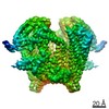
| Supplemental images |
- Downloads & links
-EMDB archive
| Map data | emd_12638.map.gz | 163.4 MB | EMDB map data format | |
|---|---|---|---|---|
| Header (meta data) | emd-12638-v30.xmlemd-12638.xml | 20.8 KB 20.8 KB | Display Display | EMDB header |
| FSC (resolution estimation) | emd_12638_fsc.xml | 13.5 KB | Display | FSC data file |
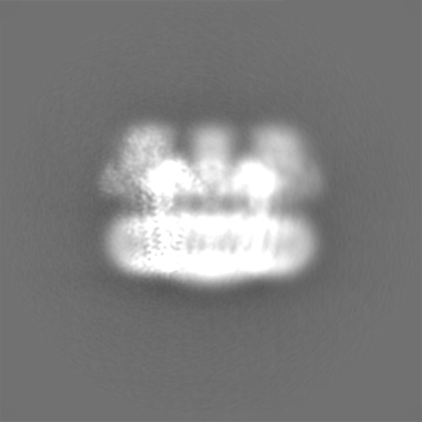
| Images |  emd_12638.png emd_12638.png | 120.7 KB | ||
| Masks | emd_12638_msk_1.map | 209.3 MB | Mask map | |
| Filedesc metadata | emd-12638.cif.gz | 6.8 KB | ||
| Others | emd_12638_half_map_1.map.gzemd_12638_half_map_2.map.gz | 164.8 MB 164.8 MB | ||
| Archive directory |  http://ftp.pdbj.org/pub/emdb/structures/EMD-12638ftp://ftp.pdbj.org/pub/emdb/structures/EMD-12638 http://ftp.pdbj.org/pub/emdb/structures/EMD-12638ftp://ftp.pdbj.org/pub/emdb/structures/EMD-12638 | HTTPS FTP |
-Related structure data
| Related structure data | 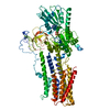 7nxfMC  7ny1C M: atomic model generated by this map C: citing same article ( |
|---|---|
| Similar structure data |

-Links
| EMDB pages | EMDB (EBI/PDBe) / EMDataResource |
|---|---|
| Related items in Molecule of the Month |


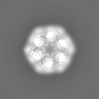
-Map
| File | Download / File: emd_12638.map.gz / Format: CCP4 / Size: 209.3 MB / Type: IMAGE STORED AS FLOATING POINT NUMBER (4 BYTES) | ||||||||||||||||||||||||||||||||||||||||||||||||||||||||||||
|---|---|---|---|---|---|---|---|---|---|---|---|---|---|---|---|---|---|---|---|---|---|---|---|---|---|---|---|---|---|---|---|---|---|---|---|---|---|---|---|---|---|---|---|---|---|---|---|---|---|---|---|---|---|---|---|---|---|---|---|---|---|
| Annotation | monomeric map derived from focused 3D classification and refinement of symmetry expanded particles | ||||||||||||||||||||||||||||||||||||||||||||||||||||||||||||
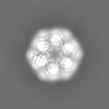
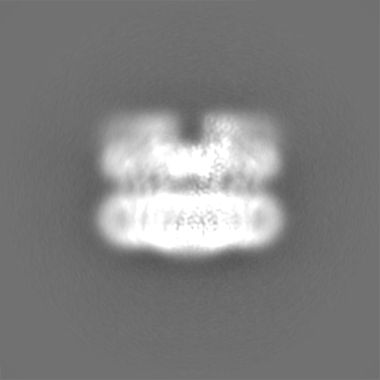
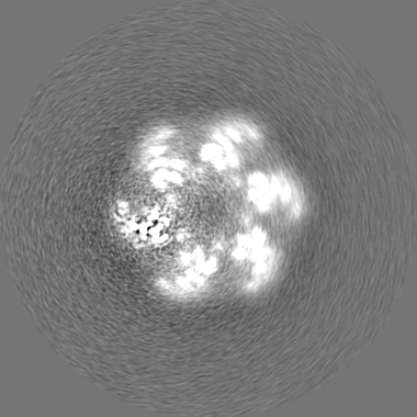
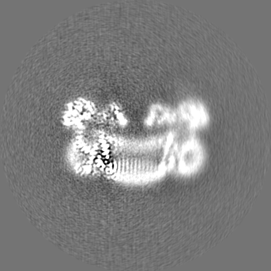
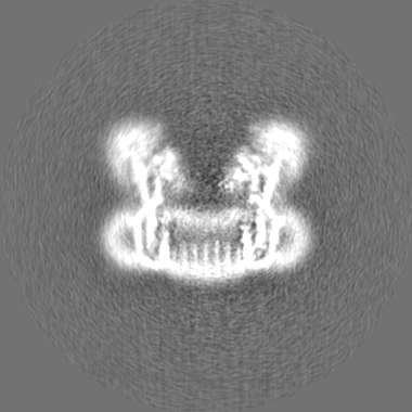
| Projections & slices | Image control
Images are generated by Spider. | ||||||||||||||||||||||||||||||||||||||||||||||||||||||||||||
| Voxel size | X=Y=Z: 0.837 Å | ||||||||||||||||||||||||||||||||||||||||||||||||||||||||||||
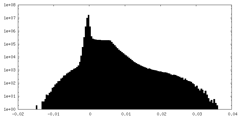
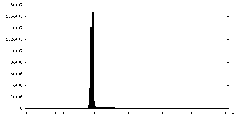
| Density |
| ||||||||||||||||||||||||||||||||||||||||||||||||||||||||||||
| Symmetry | Space group: 1 | ||||||||||||||||||||||||||||||||||||||||||||||||||||||||||||
| Details | EMDB XML:
CCP4 map header:
| ||||||||||||||||||||||||||||||||||||||||||||||||||||||||||||
 Z (Sec.)
 Y (Row.)
 X (Col.)


-Supplemental data
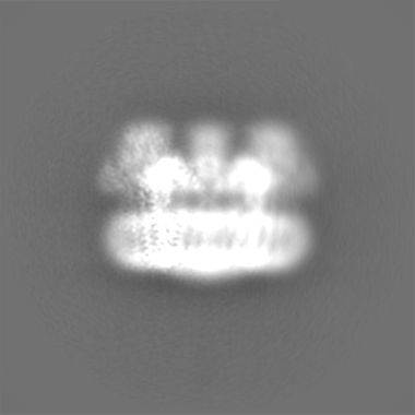
-Mask #1
| File | emd_12638_msk_1.map | ||||||||||||
|---|---|---|---|---|---|---|---|---|---|---|---|---|---|
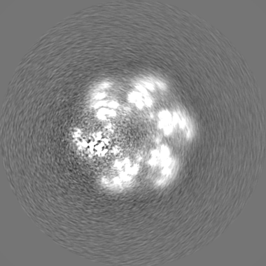
| Projections & Slices |
| ||||||||||||
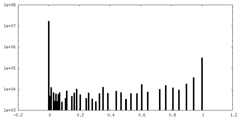
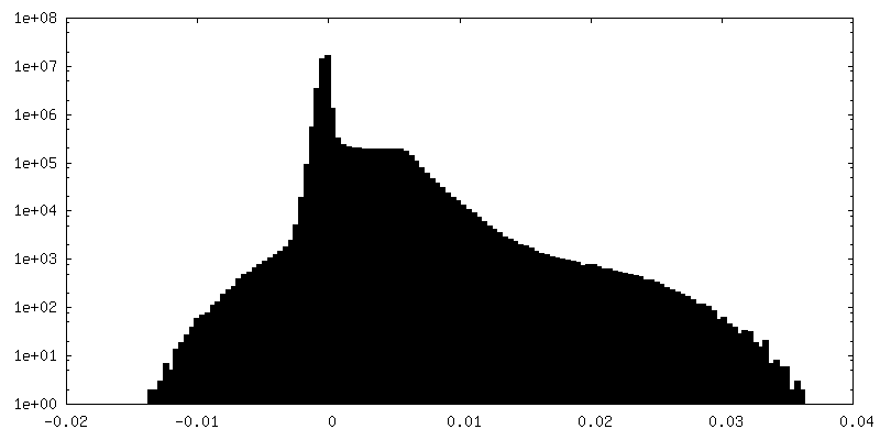
| Density Histograms |


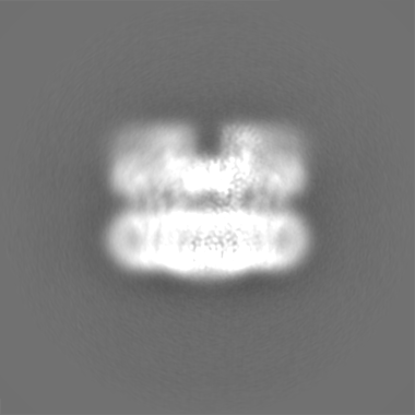
-Half map: half map 2
| File | emd_12638_half_map_1.map | ||||||||||||
|---|---|---|---|---|---|---|---|---|---|---|---|---|---|
| Annotation | half map 2 | ||||||||||||
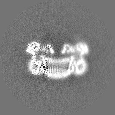
| Projections & Slices |
| ||||||||||||
| Density Histograms |


-Half map: half map 1
| File | emd_12638_half_map_2.map | ||||||||||||
|---|---|---|---|---|---|---|---|---|---|---|---|---|---|
| Annotation | half map 1 | ||||||||||||
| Projections & Slices |
| ||||||||||||
| Density Histograms |
- Sample components
-Entire : Monomeric subunit of the hexameric fungal plasma membrane proton ...
| Entire | Name: Monomeric subunit of the hexameric fungal plasma membrane proton pump in its auto-inhibited state |
|---|---|
| Components |
|
-Supramolecule #1: Monomeric subunit of the hexameric fungal plasma membrane proton ...
| Supramolecule | Name: Monomeric subunit of the hexameric fungal plasma membrane proton pump in its auto-inhibited state type: organelle_or_cellular_component / ID: 1 / Parent: 0 / Macromolecule list: #1 |
|---|---|
| Source (natural) | Organism: Neurospora crassa (fungus) / Strain: FGSC #4761 / Location in cell: plasma membrane |
-Macromolecule #1: Plasma membrane ATPase
| Macromolecule | Name: Plasma membrane ATPase / type: protein_or_peptide / ID: 1 / Number of copies: 1 / Enantiomer: LEVO / EC number: P-type H+-exporting transporter |
|---|---|
| Source (natural) | Organism: Neurospora crassa (fungus) |
| Molecular weight | Theoretical: 99.984359 KDa |
| Sequence | String: MADHSASGAP ALSTNIESGK FDEKAAEAAA YQPKPKVEDD EDEDIDALIE DLESHDGHDA EEEEEEATPG GGRVVPEDML QTDTRVGLT SEEVVQRRRK YGLNQMKEEK ENHFLKFLGF FVGPIQFVME GAAVLAAGLE DWVDFGVICG LLLLNAVVGF V QEFQAGSI ...String: MADHSASGAP ALSTNIESGK FDEKAAEAAA YQPKPKVEDD EDEDIDALIE DLESHDGHDA EEEEEEATPG GGRVVPEDML QTDTRVGLT SEEVVQRRRK YGLNQMKEEK ENHFLKFLGF FVGPIQFVME GAAVLAAGLE DWVDFGVICG LLLLNAVVGF V QEFQAGSI VDELKKTLAL KAVVLRDGTL KEIEAPEVVP GDILQVEEGT IIPADGRIVT DDAFLQVDQS ALTGESLAVD KH KGDQVFA SSAVKRGEAF VVITATGDNT FVGRAAALVN AASGGSGHFT EVLNGIGTIL LILVIFTLLI VWVSSFYRSN PIV QILEFT LAITIIGVPV GLPAVVTTTM AVGAAYLAKK KAIVQKLSAI ESLAGVEILC SDKTGTLTKN KLSLHDPYTV AGVD PEDLM LTACLAASRK KKGIDAIDKA FLKSLKYYPR AKSVLSKYKV LQFHPFDPVS KKVVAVVESP QGERITCVKG APLFV LKTV EEDHPIPEEV DQAYKNKVAE FATRGFRSLG VARKRGEGSW EILGIMPCMD PPRHDTYKTV CEAKTLGLSI KMLTGD AVG IARETSRQLG LGTNIYNAER LGLGGGGDMP GSEVYDFVEA ADGFAEVFPQ HKYNVVEILQ QRGYLVAMTG DGVNDAP SL KKADTGIAVE GSSDAARSAA DIVFLAPGLG AIIDALKTSR QIFHRMYAYV VYRIALSIHL EIFLGLWIAI LNRSLNIE L VVFIAIFADV ATLAIAYDNA PYSQTPVKWN LPKLWGMSVL LGVVLAVGTW ITVTTMYAQG ENGGIVQNFG NMDEVLFLQ ISLTENWLIF ITRANGPFWS SIPSWQLSGA IFLVDILATC FTIWGWFEHS DTSIVAVVRI WIFSFGIFCI MGGVYYILQD SVGFDNLMH GKSPKGNQKQ RSLEDFVVSL QRVSTQHEKS Q UniProtKB: Plasma membrane ATPase |
-Macromolecule #2: ADENOSINE-5'-DIPHOSPHATE
| Macromolecule | Name: ADENOSINE-5'-DIPHOSPHATE / type: ligand / ID: 2 / Number of copies: 1 / Formula: ADP |
|---|---|
| Molecular weight | Theoretical: 427.201 Da |
| Chemical component information |  ChemComp-ADP: |
-Macromolecule #3: MAGNESIUM ION
| Macromolecule | Name: MAGNESIUM ION / type: ligand / ID: 3 / Number of copies: 1 / Formula: MG |
|---|---|
| Molecular weight | Theoretical: 24.305 Da |
-Macromolecule #4: POTASSIUM ION
| Macromolecule | Name: POTASSIUM ION / type: ligand / ID: 4 / Number of copies: 1 / Formula: K |
|---|---|
| Molecular weight | Theoretical: 39.098 Da |
-Experimental details
-Structure determination
| Method | cryo EM |
|---|---|
 Processing Processing | single particle reconstruction |
| Aggregation state | particle |
-Sample preparation
| Concentration | 2 mg/mL |
|---|---|
| Buffer | pH: 6.5 |
| Grid | Model: C-flat-2/2 / Material: COPPER / Mesh: 400 / Pretreatment - Type: GLOW DISCHARGE |
| Vitrification | Cryogen name: ETHANE / Chamber humidity: 70 % / Chamber temperature: 283 K / Instrument: FEI VITROBOT MARK IV |
- Electron microscopy
| Microscope | FEI TITAN KRIOS |
|---|---|
| Image recording | Film or detector model: GATAN K3 (6k x 4k) / Average electron dose: 42.0 e/Å2 |
| Electron beam | Acceleration voltage: 300 kV / Electron source:  FIELD EMISSION GUN FIELD EMISSION GUN |
| Electron optics | Illumination mode: FLOOD BEAM / Imaging mode: BRIGHT FIELD |
| Experimental equipment |  Model: Titan Krios / Image courtesy: FEI Company |