Movie
Movie Controller


+ Open data

- Basic information
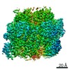
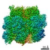
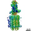
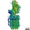
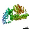
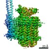
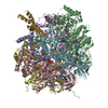
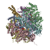
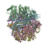
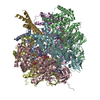
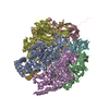
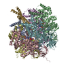
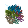
| Entry | Database: EMDB / ID: EMD-12432 | |||||||||
|---|---|---|---|---|---|---|---|---|---|---|
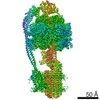
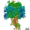
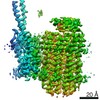
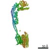
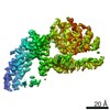
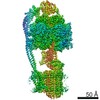
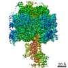
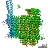
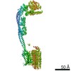
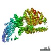
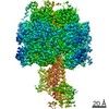
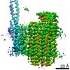
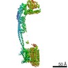
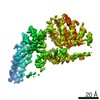
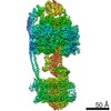
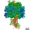
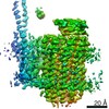
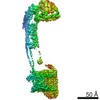
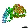
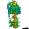
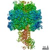
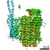
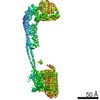
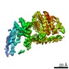
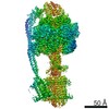
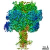
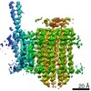
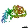
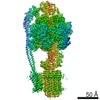
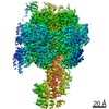
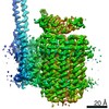
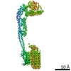
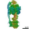
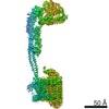
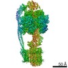
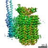
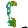
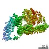
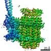
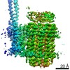
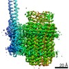
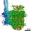
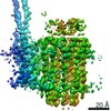
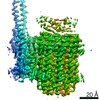
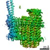
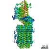
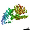
| Title | Mycobacterium smegmatis ATP synthase F1 state 1 | |||||||||
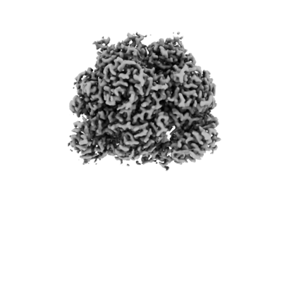
 Map data Map data | Ms F1 combiset main map | |||||||||
 Sample Sample |
| |||||||||
 Keywords Keywords | complex / synthase / HYDROLASE | |||||||||
| Function / homology |  Function and homology information Function and homology informationproton motive force-driven plasma membrane ATP synthesis / H+-transporting two-sector ATPase / proton-transporting ATP synthase complex / proton-transporting ATP synthase activity, rotational mechanism / ADP binding / ATP binding / plasma membrane Similarity search - Function | |||||||||
| Biological species |  Mycolicibacterium smegmatis MC2 155 (bacteria) / Mycolicibacterium smegmatis (strain ATCC 700084 / mc(2)155) (bacteria) / Mycobacterium smegmatis (strain ATCC 700084 / mc(2)155) (bacteria) Mycolicibacterium smegmatis MC2 155 (bacteria) / Mycolicibacterium smegmatis (strain ATCC 700084 / mc(2)155) (bacteria) / Mycobacterium smegmatis (strain ATCC 700084 / mc(2)155) (bacteria) | |||||||||
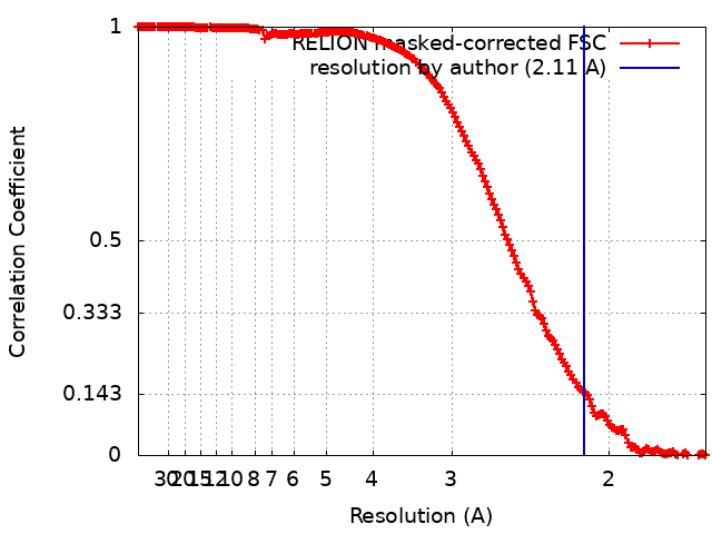
| Method | single particle reconstruction / cryo EM / Resolution: 2.11 Å | |||||||||
 Authors Authors | Montgomery MG / Petri J | |||||||||
| Funding support |  United Kingdom, 2 items United Kingdom, 2 items
| |||||||||
 Citation Citation | Journal: Proc Natl Acad Sci U S A / Year: 2021 Title: Structure of the ATP synthase from provides targets for treating tuberculosis. Authors: Martin G Montgomery / Jessica Petri / Tobias E Spikes / John E Walker / Abstract: The structure has been determined by electron cryomicroscopy of the adenosine triphosphate (ATP) synthase from This analysis confirms features in a prior description of the structure of the enzyme, ...The structure has been determined by electron cryomicroscopy of the adenosine triphosphate (ATP) synthase from This analysis confirms features in a prior description of the structure of the enzyme, but it also describes other highly significant attributes not recognized before that are crucial for understanding the mechanism and regulation of the mycobacterial enzyme. First, we resolved not only the three main states in the catalytic cycle described before but also eight substates that portray structural and mechanistic changes occurring during a 360° catalytic cycle. Second, a mechanism of auto-inhibition of ATP hydrolysis involves not only the engagement of the C-terminal region of an α-subunit in a loop in the γ-subunit, as proposed before, but also a "fail-safe" mechanism involving the b'-subunit in the peripheral stalk that enhances engagement. A third unreported characteristic is that the fused bδ-subunit contains a duplicated domain in its N-terminal region where the two copies of the domain participate in similar modes of attachment of the two of three N-terminal regions of the α-subunits. The auto-inhibitory plus the associated "fail-safe" mechanisms and the modes of attachment of the α-subunits provide targets for development of innovative antitubercular drugs. The structure also provides support for an observation made in the bovine ATP synthase that the transmembrane proton-motive force that provides the energy to drive the rotary mechanism is delivered directly and tangentially to the rotor via a Grotthuss water chain in a polar L-shaped tunnel. | |||||||||
| History |
|
- Structure visualization
| Movie |
Movie viewer |
|---|---|
| Structure viewer | EM map: SurfViewMolmilJmol/JSmol |
| Supplemental images |
- Downloads & links
-EMDB archive
| Map data | emd_12432.map.gz | 21.3 MB | EMDB map data format | |
|---|---|---|---|---|
| Header (meta data) | emd-12432-v30.xmlemd-12432.xml | 14.6 KB 14.6 KB | Display Display | EMDB header |
| FSC (resolution estimation) | emd_12432_fsc.xml | 17.6 KB | Display | FSC data file |
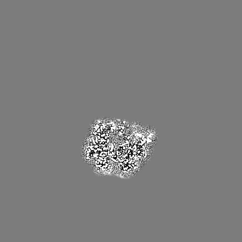
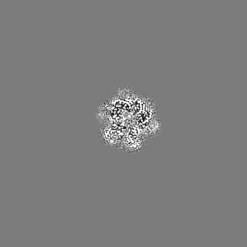
| Images |  emd_12432.png emd_12432.png | 71.2 KB | ||
| Filedesc metadata | emd-12432.cif.gz | 6.2 KB | ||
| Archive directory |  http://ftp.pdbj.org/pub/emdb/structures/EMD-12432ftp://ftp.pdbj.org/pub/emdb/structures/EMD-12432 http://ftp.pdbj.org/pub/emdb/structures/EMD-12432ftp://ftp.pdbj.org/pub/emdb/structures/EMD-12432 | HTTPS FTP |
-Related structure data
| Related structure data |  7nk7MC  7njkC 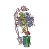 7njlC 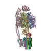 7njmC  7njnC 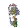 7njoC 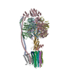 7njpC  7njqC  7njrC  7njsC  7njtC  7njuC  7njvC  7njwC 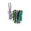 7njxC 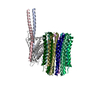 7njyC 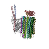 7nk9C 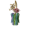 7nkbC 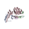 7nkdC 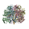 7nkhC 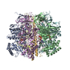 7nkjC  7nkkC 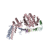 7nklC  7nknC  7nkpC 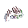 7nkqC 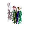 7nl9C C: citing same article ( M: atomic model generated by this map |
|---|---|
| Similar structure data |


-Links
| EMDB pages | EMDB (EBI/PDBe) / EMDataResource |
|---|---|
| Related items in Molecule of the Month |


-Map
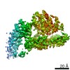
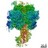
| File | Download / File: emd_12432.map.gz / Format: CCP4 / Size: 476.8 MB / Type: IMAGE STORED AS FLOATING POINT NUMBER (4 BYTES) | ||||||||||||||||||||||||||||||||||||||||||||||||||||||||||||
|---|---|---|---|---|---|---|---|---|---|---|---|---|---|---|---|---|---|---|---|---|---|---|---|---|---|---|---|---|---|---|---|---|---|---|---|---|---|---|---|---|---|---|---|---|---|---|---|---|---|---|---|---|---|---|---|---|---|---|---|---|---|
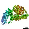
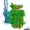
| Annotation | Ms F1 combiset main map | ||||||||||||||||||||||||||||||||||||||||||||||||||||||||||||
| Projections & slices | Image control
Images are generated by Spider. | ||||||||||||||||||||||||||||||||||||||||||||||||||||||||||||
| Voxel size | X=Y=Z: 0.83 Å | ||||||||||||||||||||||||||||||||||||||||||||||||||||||||||||
| Density |
| ||||||||||||||||||||||||||||||||||||||||||||||||||||||||||||
| Symmetry | Space group: 1 | ||||||||||||||||||||||||||||||||||||||||||||||||||||||||||||
| Details | EMDB XML:
CCP4 map header:
| ||||||||||||||||||||||||||||||||||||||||||||||||||||||||||||
 Z (Sec.)
 Y (Row.)
 X (Col.)


-Supplemental data
- Sample components
-Entire : Mycobacterium smegmatis ATP synthase
| Entire | Name: Mycobacterium smegmatis ATP synthase |
|---|---|
| Components |
|
-Supramolecule #1: Mycobacterium smegmatis ATP synthase
| Supramolecule | Name: Mycobacterium smegmatis ATP synthase / type: complex / ID: 1 / Parent: 0 / Macromolecule list: #1-#3 |
|---|---|
| Source (natural) | Organism: Mycolicibacterium smegmatis MC2 155 (bacteria) |
| Molecular weight | Theoretical: 547 KDa |
-Macromolecule #1: ATP synthase subunit alpha
| Macromolecule | Name: ATP synthase subunit alpha / type: protein_or_peptide / ID: 1 / Number of copies: 3 / Enantiomer: LEVO / EC number: H+-transporting two-sector ATPase |
|---|---|
| Source (natural) | Organism: Mycolicibacterium smegmatis (strain ATCC 700084 / mc(2)155) (bacteria) Strain: ATCC 700084 / mc(2)155 |
| Molecular weight | Theoretical: 58.951461 KDa |
| Recombinant expression | Organism: Mycolicibacterium smegmatis (bacteria) |
| Sequence | String: MAELTISAAD IEGAIEDYVS SFSADTEREE IGTVIDAGDG IAHVEGLPSV MTQELLEFPG GVLGVALNLD EHSVGAVILG EFEKIEEGQ QVKRTGEVLS VPVGDAFLGR VVNPLGQPID GQGDIAAETR RALELQAPSV VQRQSVSEPL QTGIKAIDAM T PIGRGQRQ ...String: MAELTISAAD IEGAIEDYVS SFSADTEREE IGTVIDAGDG IAHVEGLPSV MTQELLEFPG GVLGVALNLD EHSVGAVILG EFEKIEEGQ QVKRTGEVLS VPVGDAFLGR VVNPLGQPID GQGDIAAETR RALELQAPSV VQRQSVSEPL QTGIKAIDAM T PIGRGQRQ LIIGDRKTGK TAVCVDTILN QREAWLTGDP KQQVRCVYVA IGQKGTTIAS VKRALEEGGA MEYTTIVAAP AS DAAGFKW LAPYTGSAIG QHWMYNGKHV LIVFDDLSKQ ADAYRAISLL LRRPPGREAF PGDVFYLHSR LLERCAKLSD ELG GGSMTG LPIIETKAND ISAFIPTNVI SITDGQCFLE SDLFNQGVRP AINVGVSVSR VGGAAQIKAM KEVAGSLRLD LSQY RELEA FAAFASDLDA ASKAQLDRGA RLVELLKQPQ YSPLAVEEQV VAIFLGTQGH LDSVPVEDVQ RFESELLEHV KASHS DIFD GIRETKKLSE EAEEKLVSVI NEFKKGFQAS DGSSVVVSEN AEALDPEDLE KESVKVRKPA PKKA UniProtKB: ATP synthase subunit alpha |
-Macromolecule #2: ATP synthase subunit beta
| Macromolecule | Name: ATP synthase subunit beta / type: protein_or_peptide / ID: 2 / Details: Uniprot A0R200. The crossref text box is blank. / Number of copies: 3 / Enantiomer: LEVO / EC number: H+-transporting two-sector ATPase |
|---|---|
| Source (natural) | Organism: Mycolicibacterium smegmatis (strain ATCC 700084 / mc(2)155) (bacteria) Strain: ATCC 700084 / mc(2)155 |
| Molecular weight | Theoretical: 51.670453 KDa |
| Recombinant expression | Organism: Mycolicibacterium smegmatis (bacteria) |
| Sequence | String: MTATAEKTAG RVVRITGPVV DVEFPRGSVP ELFNALHAEI TFGALAKTLT LEVAQHLGDS LVRCISMQPT DGLVRGVEVT DTGASISVP VGDGVKGHVF NALGDCLDDP GYGKDFEHWS IHRKPPAFSD LEPRTEMLET GLKVVDLLTP YVRGGKIALF G GAGVGKTV ...String: MTATAEKTAG RVVRITGPVV DVEFPRGSVP ELFNALHAEI TFGALAKTLT LEVAQHLGDS LVRCISMQPT DGLVRGVEVT DTGASISVP VGDGVKGHVF NALGDCLDDP GYGKDFEHWS IHRKPPAFSD LEPRTEMLET GLKVVDLLTP YVRGGKIALF G GAGVGKTV LIQEMINRIA RNFGGTSVFA GVGERTREGN DLWVELADAN VLKDTALVFG QMDEPPGTRM RVALSALTMA EF FRDEQGQ DVLLFIDNIF RFTQAGSEVS TLLGRMPSAV GYQPTLADEM GELQERITST RGRSITSMQA VYVPADDYTD PAP ATTFAH LDATTELSRA VFSKGIFPAV DPLASSSTIL DPAIVGDEHY RVAQEVIRIL QRYKDLQDII AILGIDELSE EDKQ LVNRA RRIERFLSQN MMAAEQFTGQ PGSTVPLKET IEAFDKLTKG EFDHLPEQAF FLIGGLDDLA KKAESLGAKL UniProtKB: ATP synthase subunit beta |
-Macromolecule #3: ATP synthase gamma chain
| Macromolecule | Name: ATP synthase gamma chain / type: protein_or_peptide / ID: 3 Details: This alignment is not correct. There are no mismatches but there are breaks in the peptide. UNIPROT A0R201. The crossref text box is missing below. Number of copies: 1 / Enantiomer: LEVO |
|---|---|
| Source (natural) | Organism: Mycobacterium smegmatis (strain ATCC 700084 / mc(2)155) (bacteria) Strain: ATCC 700084 / mc(2)155 |
| Molecular weight | Theoretical: 33.439836 KDa |
| Recombinant expression | Organism: Mycolicibacterium smegmatis (bacteria) |
| Sequence | String: MAATLRELRG RIRSAGSIKK ITKAQELIAT SRIAKAQARV EAARPYAAEI TNMLTELAGA SALDHPLLVE RKQPKRAGVL VVSSDRGLC GAYNANVLRR AEELFSLLRD EGKDPVLYVV GRKALGYFSF RQRTVVESWT GFSERPTYEN AREIADTLVN A FMAGADDE ...String: MAATLRELRG RIRSAGSIKK ITKAQELIAT SRIAKAQARV EAARPYAAEI TNMLTELAGA SALDHPLLVE RKQPKRAGVL VVSSDRGLC GAYNANVLRR AEELFSLLRD EGKDPVLYVV GRKALGYFSF RQRTVVESWT GFSERPTYEN AREIADTLVN A FMAGADDE GDDAGADGIL GVDELHIVFT EFRSMLSQTA VARRAAPMEV EYVGEVETGP RTLYSFEPDP ETLFDALLPR YI ATRVYAA LLEAAASESA SRRRAMKSAT DNADDLIKAL TLAANRERQA QITQEISEIV GGANALAGSK UniProtKB: ATP synthase gamma chain |
-Macromolecule #4: ADENOSINE-5'-TRIPHOSPHATE
| Macromolecule | Name: ADENOSINE-5'-TRIPHOSPHATE / type: ligand / ID: 4 / Number of copies: 4 / Formula: ATP |
|---|---|
| Molecular weight | Theoretical: 507.181 Da |
| Chemical component information |  ChemComp-ATP: |
-Macromolecule #5: MAGNESIUM ION
| Macromolecule | Name: MAGNESIUM ION / type: ligand / ID: 5 / Number of copies: 5 / Formula: MG |
|---|---|
| Molecular weight | Theoretical: 24.305 Da |
-Macromolecule #6: ADENOSINE-5'-DIPHOSPHATE
| Macromolecule | Name: ADENOSINE-5'-DIPHOSPHATE / type: ligand / ID: 6 / Number of copies: 2 / Formula: ADP |
|---|---|
| Molecular weight | Theoretical: 427.201 Da |
| Chemical component information |  ChemComp-ADP: |
-Macromolecule #7: water
| Macromolecule | Name: water / type: ligand / ID: 7 / Number of copies: 459 / Formula: HOH |
|---|---|
| Molecular weight | Theoretical: 18.015 Da |
| Chemical component information |  ChemComp-HOH: |
-Experimental details
-Structure determination
| Method | cryo EM |
|---|---|
 Processing Processing | single particle reconstruction |
| Aggregation state | particle |
-Sample preparation
| Buffer | pH: 8 |
|---|---|
| Vitrification | Cryogen name: ETHANE |
- Electron microscopy
| Microscope | FEI TITAN KRIOS |
|---|---|
| Image recording | Film or detector model: GATAN K3 (6k x 4k) / Average electron dose: 59.86 e/Å2 |
| Electron beam | Acceleration voltage: 300 kV / Electron source:  FIELD EMISSION GUN FIELD EMISSION GUN |
| Electron optics | Illumination mode: FLOOD BEAM / Imaging mode: BRIGHT FIELD |
| Experimental equipment |  Model: Titan Krios / Image courtesy: FEI Company |