ムービー
ムービー コントローラー


+ データを開く

- 基本情報
基本情報
| 登録情報 | データベース: EMDB / ID: EMD-12334 | |||||||||||||||||||||
|---|---|---|---|---|---|---|---|---|---|---|---|---|---|---|---|---|---|---|---|---|---|---|
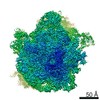
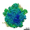
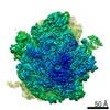
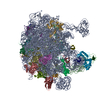
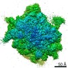
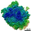
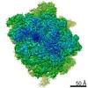
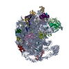
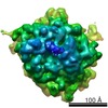
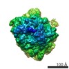
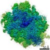
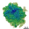
| タイトル | VgaL, an antibiotic resistance ABCF, in complex with 70S ribosome from Listeria monocytogenes | |||||||||||||||||||||
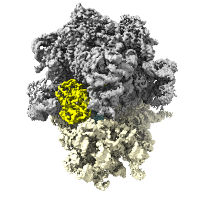
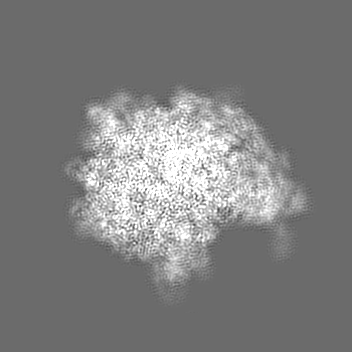
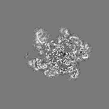
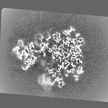
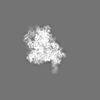
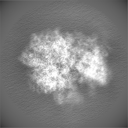
 マップデータ マップデータ | VgaL in complex with 70S ribosome from Listeria monocytogenes. Post-processed with automatically estimated B-factor. | |||||||||||||||||||||
 試料 試料 |
| |||||||||||||||||||||
 キーワード キーワード | Antibiotic resistance element / ABCF / antibiotic resistance / ATPase / protein synthesis / RIBOSOME | |||||||||||||||||||||
| 機能・相同性 |  機能・相同性情報 機能・相同性情報positive regulation of translational fidelity / ATPase-coupled transmembrane transporter activity / maturation of SSU-rRNA from tricistronic rRNA transcript (SSU-rRNA, 5.8S rRNA, LSU-rRNA) / mRNA 5'-UTR binding / large ribosomal subunit / ribosomal small subunit assembly / small ribosomal subunit / small ribosomal subunit rRNA binding / transferase activity / 5S rRNA binding ...positive regulation of translational fidelity / ATPase-coupled transmembrane transporter activity / maturation of SSU-rRNA from tricistronic rRNA transcript (SSU-rRNA, 5.8S rRNA, LSU-rRNA) / mRNA 5'-UTR binding / large ribosomal subunit / ribosomal small subunit assembly / small ribosomal subunit / small ribosomal subunit rRNA binding / transferase activity / 5S rRNA binding / large ribosomal subunit rRNA binding / cytosolic small ribosomal subunit / cytosolic large ribosomal subunit / tRNA binding / cytoplasmic translation / rRNA binding / negative regulation of translation / ribosome / structural constituent of ribosome / ribonucleoprotein complex / translation / mRNA binding / RNA binding / zinc ion binding / ATP binding / cytoplasm / cytosol 類似検索 - 分子機能 | |||||||||||||||||||||
| 生物種 |  Listeria monocytogenes EGD-e (バクテリア) / Listeria monocytogenes EGD-E (バクテリア) Listeria monocytogenes EGD-e (バクテリア) / Listeria monocytogenes EGD-E (バクテリア) | |||||||||||||||||||||
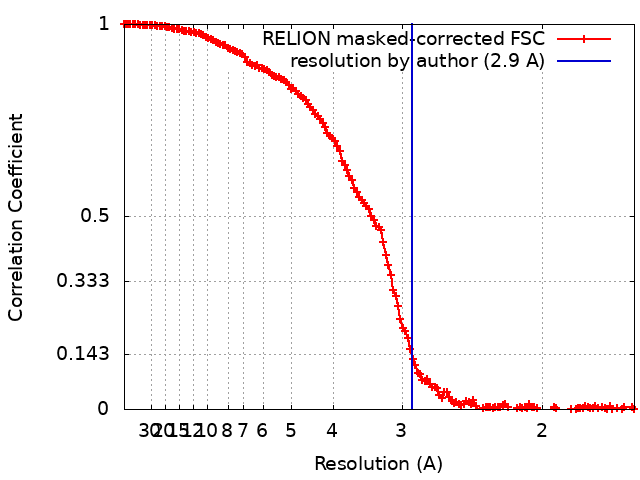
| 手法 | 単粒子再構成法 / クライオ電子顕微鏡法 / 解像度: 2.9 Å | |||||||||||||||||||||
 データ登録者 データ登録者 | Crowe-McAuliffe C / Turnbull KJ | |||||||||||||||||||||
| 資金援助 |  ドイツ, ドイツ,  スウェーデン, スウェーデン,  Estonia, 6件 Estonia, 6件
| |||||||||||||||||||||
 引用 引用 | ジャーナル: Nat Commun / 年: 2021 タイトル: Structural basis of ABCF-mediated resistance to pleuromutilin, lincosamide, and streptogramin A antibiotics in Gram-positive pathogens. 著者: Caillan Crowe-McAuliffe / Victoriia Murina / Kathryn Jane Turnbull / Marje Kasari / Merianne Mohamad / Christine Polte / Hiraku Takada / Karolis Vaitkevicius / Jörgen Johansson / Zoya ...著者: Caillan Crowe-McAuliffe / Victoriia Murina / Kathryn Jane Turnbull / Marje Kasari / Merianne Mohamad / Christine Polte / Hiraku Takada / Karolis Vaitkevicius / Jörgen Johansson / Zoya Ignatova / Gemma C Atkinson / Alex J O'Neill / Vasili Hauryliuk / Daniel N Wilson /  要旨: Target protection proteins confer resistance to the host organism by directly binding to the antibiotic target. One class of such proteins are the antibiotic resistance (ARE) ATP-binding cassette ...Target protection proteins confer resistance to the host organism by directly binding to the antibiotic target. One class of such proteins are the antibiotic resistance (ARE) ATP-binding cassette (ABC) proteins of the F-subtype (ARE-ABCFs), which are widely distributed throughout Gram-positive bacteria and bind the ribosome to alleviate translational inhibition from antibiotics that target the large ribosomal subunit. Here, we present single-particle cryo-EM structures of ARE-ABCF-ribosome complexes from three Gram-positive pathogens: Enterococcus faecalis LsaA, Staphylococcus haemolyticus VgaA and Listeria monocytogenes VgaL. Supported by extensive mutagenesis analysis, these structures enable a general model for antibiotic resistance mediated by these ARE-ABCFs to be proposed. In this model, ABCF binding to the antibiotic-stalled ribosome mediates antibiotic release via mechanistically diverse long-range conformational relays that converge on a few conserved ribosomal RNA nucleotides located at the peptidyltransferase center. These insights are important for the future development of antibiotics that overcome such target protection resistance mechanisms. | |||||||||||||||||||||
| 履歴 |
|
- 構造の表示
構造の表示
| ムービー |
ムービービューア |
|---|---|
| 構造ビューア | EMマップ: SurfViewMolmilJmol/JSmol |
| 添付画像 |
- ダウンロードとリンク
-EMDBアーカイブ
| マップデータ | emd_12334.map.gz | 20.1 MB | EMDBマップデータ形式 | |
|---|---|---|---|---|
| ヘッダ (付随情報) | emd-12334-v30.xmlemd-12334.xml | 81.5 KB 81.5 KB | 表示 表示 | EMDBヘッダ |
| FSC (解像度算出) | emd_12334_fsc.xml | 14.9 KB | 表示 | FSCデータファイル |
| 画像 |  emd_12334.png emd_12334.png | 162.4 KB | ||
| Filedesc metadata | emd-12334.cif.gz | 14.5 KB | ||
| その他 | emd_12334_additional_1.map.gzemd_12334_additional_2.map.gzemd_12334_additional_3.map.gzemd_12334_additional_4.map.gzemd_12334_additional_5.map.gzemd_12334_half_map_1.map.gzemd_12334_half_map_2.map.gz | 72.1 MB 18.8 MB 9.2 MB 3.4 MB 5.7 MB 226.4 MB 226.5 MB | ||
| アーカイブディレクトリ |  http://ftp.pdbj.org/pub/emdb/structures/EMD-12334ftp://ftp.pdbj.org/pub/emdb/structures/EMD-12334 http://ftp.pdbj.org/pub/emdb/structures/EMD-12334ftp://ftp.pdbj.org/pub/emdb/structures/EMD-12334 | HTTPS FTP |
-検証レポート
| 文書・要旨 | emd_12334_validation.pdf.gz | 1 MB | 表示 | EMDB検証レポート |
|---|---|---|---|---|
| 文書・詳細版 | emd_12334_full_validation.pdf.gz | 1 MB | 表示 | |
| XML形式データ | emd_12334_validation.xml.gz | 22.7 KB | 表示 | |
| CIF形式データ | emd_12334_validation.cif.gz | 28.8 KB | 表示 | |
| アーカイブディレクトリ | https://ftp.pdbj.org/pub/emdb/validation_reports/EMD-12334ftp://ftp.pdbj.org/pub/emdb/validation_reports/EMD-12334 | HTTPS FTP |
-関連構造データ
| 関連構造データ |  7nhnMC  7nhkC 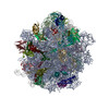 7nhlC 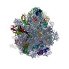 7nhmC M: このマップから作成された原子モデル C: 同じ文献を引用 ( |
|---|---|
| 類似構造データ | |
| 電子顕微鏡画像生データ | EMPIAR-10684 (タイトル: Affinity-purified VgaL in complex with 70S ribosomes from Listeria monocytogenes Data size: 146.0 Data #1: Unaligned multi-frame micrographs of VgaL bound to 70S ribosome from Listeria monocytogenes [micrographs - multiframe]) |

-リンク
| EMDBのページ | EMDB (EBI/PDBe) / EMDataResource |
|---|---|
| 「今月の分子」の関連する項目 |


-マップ
| ファイル | ダウンロード / ファイル: emd_12334.map.gz / 形式: CCP4 / 大きさ: 166.4 MB / タイプ: IMAGE STORED AS FLOATING POINT NUMBER (4 BYTES) | ||||||||||||||||||||||||||||||||||||||||||||||||||||||||||||
|---|---|---|---|---|---|---|---|---|---|---|---|---|---|---|---|---|---|---|---|---|---|---|---|---|---|---|---|---|---|---|---|---|---|---|---|---|---|---|---|---|---|---|---|---|---|---|---|---|---|---|---|---|---|---|---|---|---|---|---|---|---|
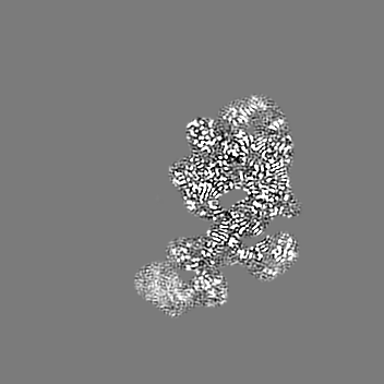
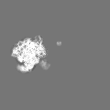
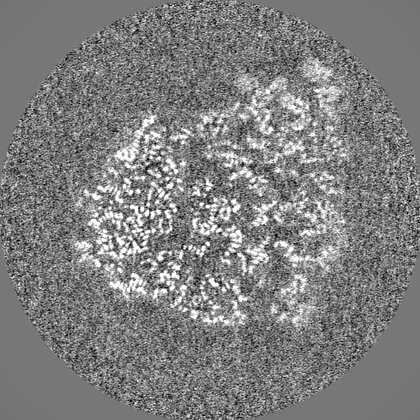
| 注釈 | VgaL in complex with 70S ribosome from Listeria monocytogenes. Post-processed with automatically estimated B-factor. | ||||||||||||||||||||||||||||||||||||||||||||||||||||||||||||
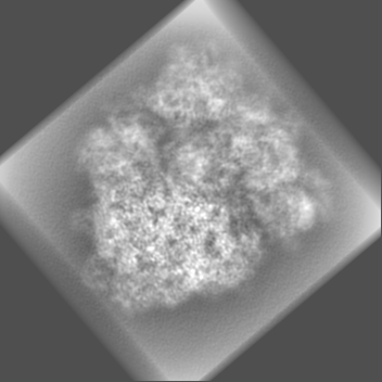
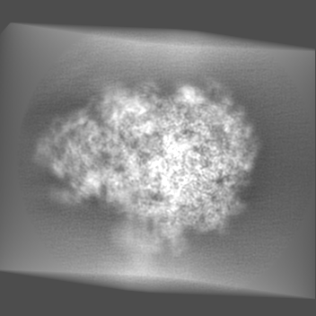
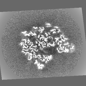
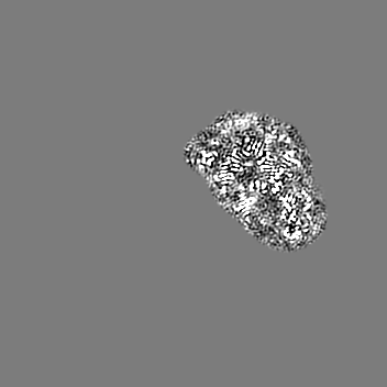
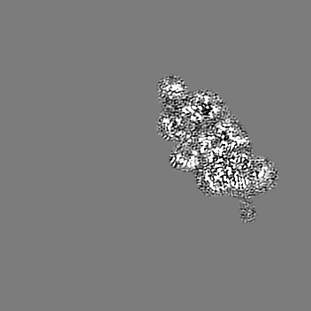
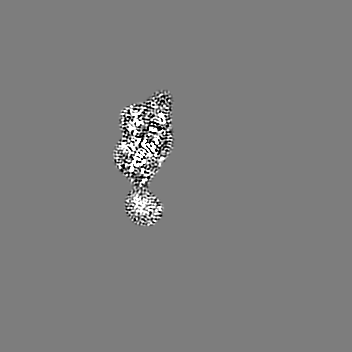
| 投影像・断面図 | 画像のコントロール
画像は Spider により作成 | ||||||||||||||||||||||||||||||||||||||||||||||||||||||||||||
| ボクセルのサイズ | X=Y=Z: 1.041 Å | ||||||||||||||||||||||||||||||||||||||||||||||||||||||||||||
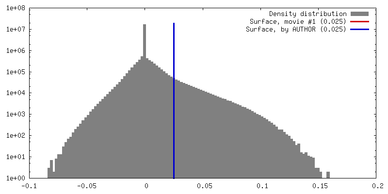
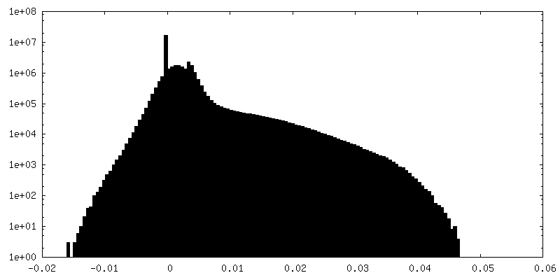
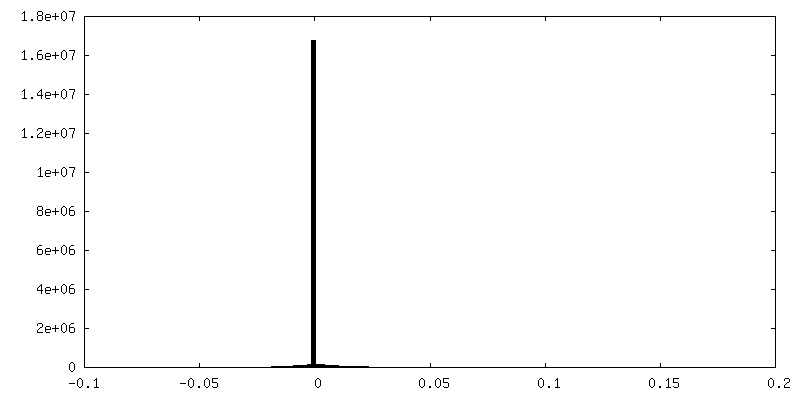
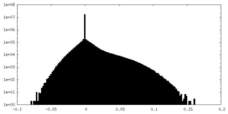
| 密度 |
| ||||||||||||||||||||||||||||||||||||||||||||||||||||||||||||
| 対称性 | 空間群: 1 | ||||||||||||||||||||||||||||||||||||||||||||||||||||||||||||
| 詳細 | EMDB XML:
CCP4マップ ヘッダ情報:
| ||||||||||||||||||||||||||||||||||||||||||||||||||||||||||||
 Z (Sec.)
 Y (Row.)
 X (Col.)


-添付データ
-追加マップ: VgaL in complex with 70S ribosome from Listeria...
| ファイル | emd_12334_additional_1.map | ||||||||||||
|---|---|---|---|---|---|---|---|---|---|---|---|---|---|
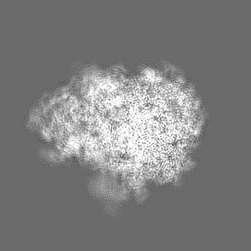
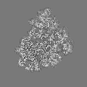
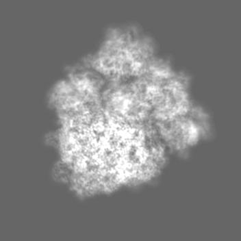
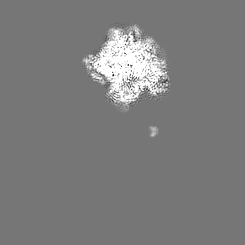
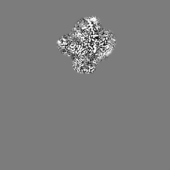
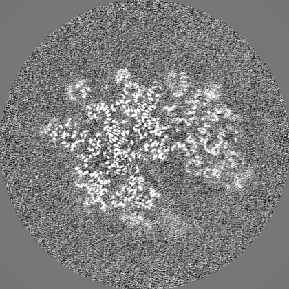
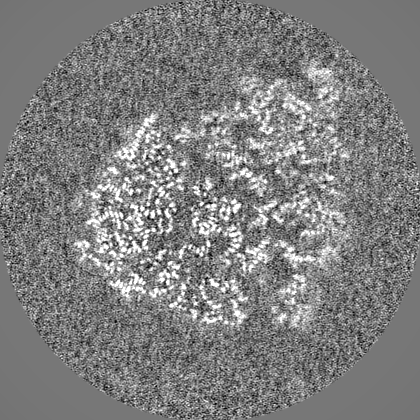
| 注釈 | VgaL in complex with 70S ribosome from Listeria monocytogenes. Output of RELION Refine3D. | ||||||||||||
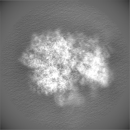
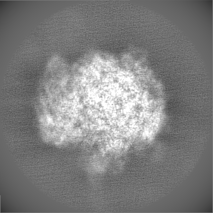
| 投影像・断面図 |
| ||||||||||||
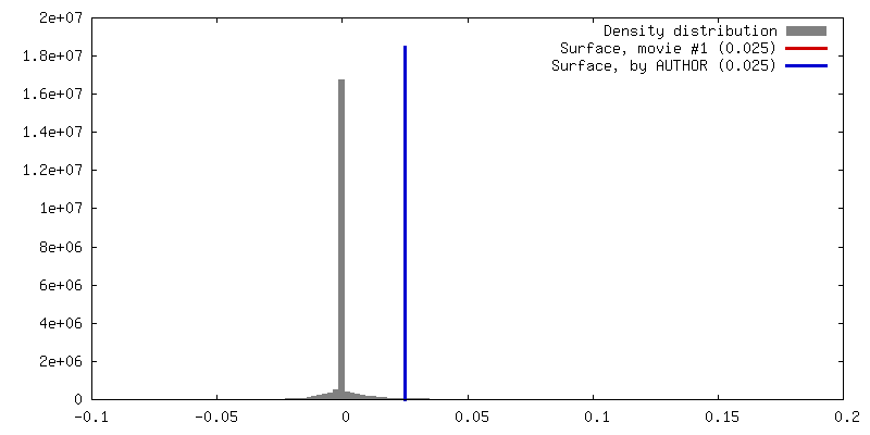
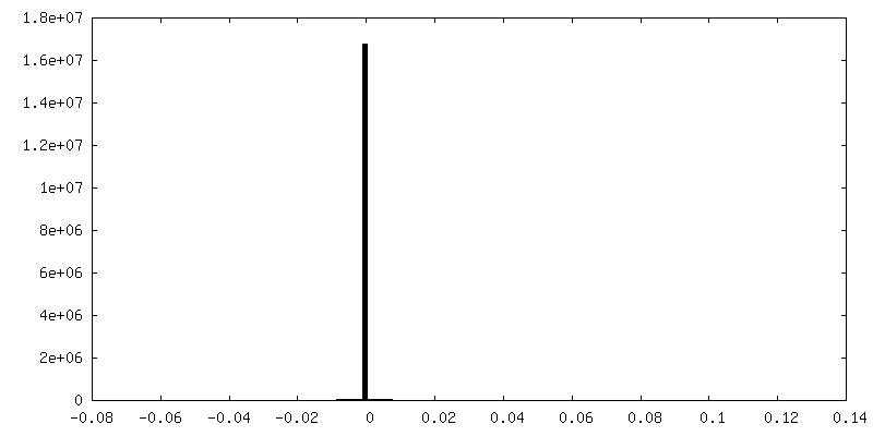
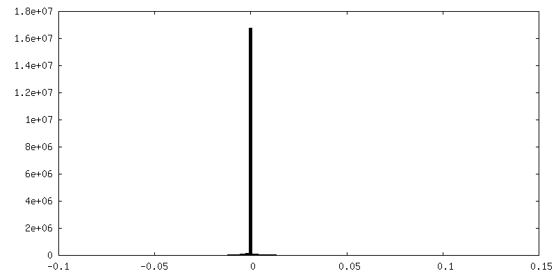
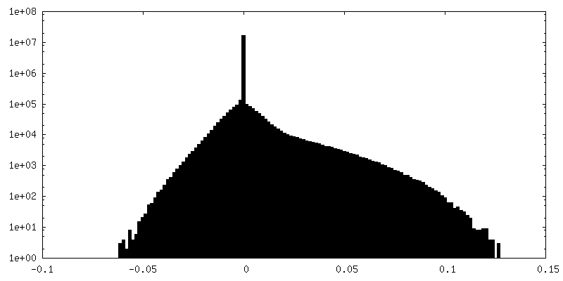
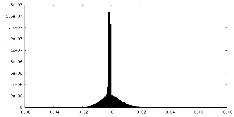
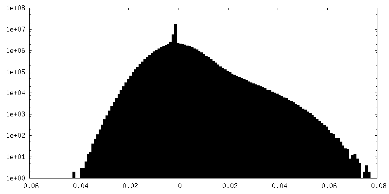
| 密度ヒストグラム |


-追加マップ: VgaL in complex with 70S ribosome from Listeria...
| ファイル | emd_12334_additional_2.map | ||||||||||||
|---|---|---|---|---|---|---|---|---|---|---|---|---|---|
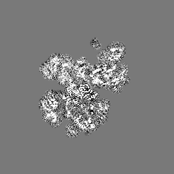
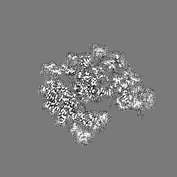
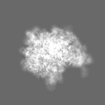
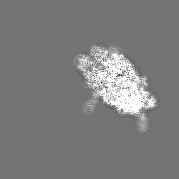
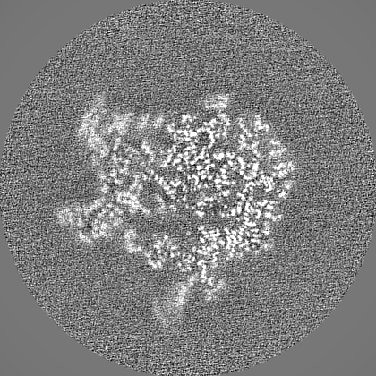
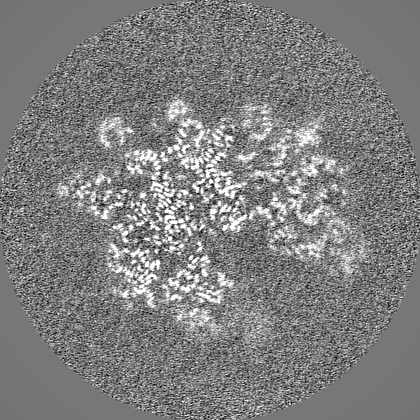
| 注釈 | VgaL in complex with 70S ribosome from Listeria monocytogenes. Filtered by local resolution. | ||||||||||||
| 投影像・断面図 |
| ||||||||||||
| 密度ヒストグラム |


-追加マップ: Multibody refinement of large ribosomal subunit and vgaL NBDs.
| ファイル | emd_12334_additional_3.map | ||||||||||||
|---|---|---|---|---|---|---|---|---|---|---|---|---|---|
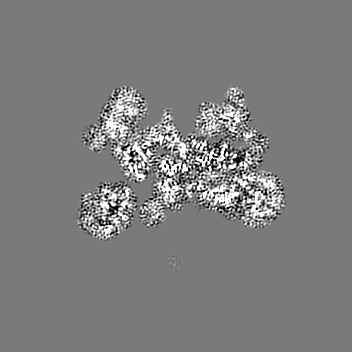
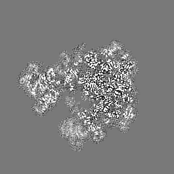
| 注釈 | Multibody refinement of large ribosomal subunit and vgaL NBDs. | ||||||||||||
| 投影像・断面図 |
| ||||||||||||
| 密度ヒストグラム |

-追加マップ: Multibody refinement of small ribosomal subunit head.
| ファイル | emd_12334_additional_4.map | ||||||||||||
|---|---|---|---|---|---|---|---|---|---|---|---|---|---|
| 注釈 | Multibody refinement of small ribosomal subunit head. | ||||||||||||
| 投影像・断面図 |
| ||||||||||||
| 密度ヒストグラム |


-追加マップ: Multibody refinement of small ribosomal subunit body.
| ファイル | emd_12334_additional_5.map | ||||||||||||
|---|---|---|---|---|---|---|---|---|---|---|---|---|---|
| 注釈 | Multibody refinement of small ribosomal subunit body. | ||||||||||||
| 投影像・断面図 |
| ||||||||||||
| 密度ヒストグラム |

-ハーフマップ: VgaL in complex with 70S ribosome from Listeria...
| ファイル | emd_12334_half_map_1.map | ||||||||||||
|---|---|---|---|---|---|---|---|---|---|---|---|---|---|
| 注釈 | VgaL in complex with 70S ribosome from Listeria monocytogenes. Half map 1. | ||||||||||||
| 投影像・断面図 |
| ||||||||||||
| 密度ヒストグラム |


-ハーフマップ: VgaL in complex with 70S ribosome from Listeria...
| ファイル | emd_12334_half_map_2.map | ||||||||||||
|---|---|---|---|---|---|---|---|---|---|---|---|---|---|
| 注釈 | VgaL in complex with 70S ribosome from Listeria monocytogenes. Half map 2. | ||||||||||||
| 投影像・断面図 |
| ||||||||||||
| 密度ヒストグラム |


- 試料の構成要素
+全体 : VgaL in complex with 70S ribosome, mRNA, and distorted P-tRNA fro...
+超分子 #1: VgaL in complex with 70S ribosome, mRNA, and distorted P-tRNA fro...
+分子 #1: Lmo0919 protein
+分子 #6: 50S ribosomal protein L2
+分子 #7: 50S ribosomal protein L3
+分子 #8: 50S ribosomal protein L4
+分子 #9: 50S ribosomal protein L5
+分子 #10: 50S ribosomal protein L6
+分子 #11: 50S ribosomal protein L13
+分子 #12: 50S ribosomal protein L14
+分子 #13: 50S ribosomal protein L15
+分子 #14: 50S ribosomal protein L16
+分子 #15: 50S ribosomal protein L17
+分子 #16: 50S ribosomal protein L18
+分子 #17: 50S ribosomal protein L19
+分子 #18: 50S ribosomal protein L20
+分子 #19: 50S ribosomal protein L21
+分子 #20: 50S ribosomal protein L22
+分子 #21: 50S ribosomal protein L23
+分子 #22: 50S ribosomal protein L24
+分子 #23: 50S ribosomal protein L27
+分子 #24: 50S ribosomal protein L28
+分子 #25: 50S ribosomal protein L29
+分子 #26: 50S ribosomal protein L30
+分子 #27: 50S ribosomal protein L32-2
+分子 #28: 50S ribosomal protein L33 1
+分子 #29: 50S ribosomal protein L34
+分子 #30: 50S ribosomal protein L35
+分子 #31: 50S ribosomal protein L36
+分子 #33: 30S ribosomal protein S2
+分子 #34: 30S ribosomal protein S3
+分子 #35: 30S ribosomal protein S4
+分子 #36: 30S ribosomal protein S5
+分子 #37: 30S ribosomal protein S6
+分子 #38: 30S ribosomal protein S7
+分子 #39: 30S ribosomal protein S8
+分子 #40: 30S ribosomal protein S9
+分子 #41: 30S ribosomal protein S10
+分子 #42: 30S ribosomal protein S11
+分子 #43: 30S ribosomal protein S12
+分子 #44: 30S ribosomal protein S13
+分子 #45: 30S ribosomal protein S14 type Z
+分子 #46: 30S ribosomal protein S15
+分子 #47: 30S ribosomal protein S16
+分子 #48: 30S ribosomal protein S17
+分子 #49: 30S ribosomal protein S18
+分子 #50: 30S ribosomal protein S19
+分子 #51: 30S ribosomal protein S20
+分子 #52: 50S ribosomal protein L31 type B
+分子 #2: tRNA-fMet
+分子 #3: RNA (5'-R(P*GP*AP*GP*GP*UP*NP*NP*NP*NP*NP*NP*AP*UP*G)-3')
+分子 #4: 23S rRNA
+分子 #5: 16S rRNA
+分子 #32: 5S rRNA
+分子 #53: ADENOSINE-5'-TRIPHOSPHATE

+分子 #54: MAGNESIUM ION
+分子 #55: SPERMIDINE

+分子 #56: POTASSIUM ION
+分子 #57: ZINC ION
-実験情報
-構造解析
| 手法 | クライオ電子顕微鏡法 |
|---|---|
 解析 解析 | 単粒子再構成法 |
| 試料の集合状態 | particle |
-試料調製
| 緩衝液 | pH: 7.5 |
|---|---|
| 凍結 | 凍結剤: ETHANE / チャンバー内湿度: 100 % / チャンバー内温度: 277.15 K / 装置: FEI VITROBOT MARK III |
- 電子顕微鏡法
| 顕微鏡 | FEI TITAN KRIOS |
|---|---|
| 撮影 | フィルム・検出器のモデル: GATAN K2 SUMMIT (4k x 4k) 撮影したグリッド数: 1 / 平均電子線量: 26.3 e/Å2 |
| 電子線 | 加速電圧: 300 kV / 電子線源:  FIELD EMISSION GUN FIELD EMISSION GUN |
| 電子光学系 | 照射モード: FLOOD BEAM / 撮影モード: BRIGHT FIELD / Cs: 2.7 mm 最大 デフォーカス(公称値): -1.9000000000000001 µm 最小 デフォーカス(公称値): -0.7000000000000001 µm 倍率(公称値): 165000 |
| 実験機器 |  モデル: Titan Krios / 画像提供: FEI Company |