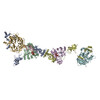
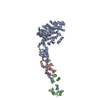
Journal: Nucleic Acids Res / Year: 2020 Title: The topology of chromatin-binding domains in the NuRD deacetylase complex. Authors: Christopher J Millard / Louise Fairall / Timothy J Ragan / Christos G Savva / John W R Schwabe / Abstract: Class I histone deacetylase complexes play essential roles in many nuclear processes. Whilst they contain a common catalytic subunit, they have diverse modes of action determined by associated ...Class I histone deacetylase complexes play essential roles in many nuclear processes. Whilst they contain a common catalytic subunit, they have diverse modes of action determined by associated factors in the distinct complexes. The deacetylase module from the NuRD complex contains three protein domains that control the recruitment of chromatin to the deacetylase enzyme, HDAC1/2. Using biochemical approaches and cryo-electron microscopy, we have determined how three chromatin-binding domains (MTA1-BAH, MBD2/3 and RBBP4/7) are assembled in relation to the core complex so as to facilitate interaction of the complex with the genome. We observe a striking arrangement of the BAH domains suggesting a potential mechanism for binding to di-nucleosomes. We also find that the WD40 domains from RBBP4 are linked to the core with surprising flexibility that is likely important for chromatin engagement. A single MBD2 protein binds asymmetrically to the dimerisation interface of the complex. This symmetry mismatch explains the stoichiometry of the complex. Finally, our structures suggest how the holo-NuRD might assemble on a di-nucleosome substrate.
History
Deposition
Oct 14, 2020
-
Header (metadata) release
Nov 11, 2020
-
Map release
Nov 11, 2020
-
Update
May 1, 2024
-
Current status
May 1, 2024
Processing site: PDBe / Status: Released
-
Structure visualization
Movie
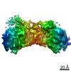
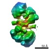
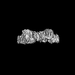
Surface view with section colored by density value
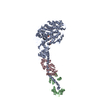
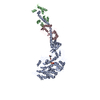
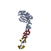
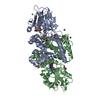
Entire : NuRD deacetylase complex containing two copies of MTA1 and HDAC1 ...
Entire
Name: NuRD deacetylase complex containing two copies of MTA1 and HDAC1 and a single copy of MBD2
Components
Complex: NuRD deacetylase complex containing two copies of MTA1 and HDAC1 and a single copy of MBD2
Protein or peptide: Methyl-CpG-binding domain protein 2
Protein or peptide: Metastasis-associated protein MTA1
Protein or peptide: Histone deacetylase 1
Ligand: INOSITOL HEXAKISPHOSPHATE
Ligand: ZINC ION
Ligand: POTASSIUM ION
-
Supramolecule #1: NuRD deacetylase complex containing two copies of MTA1 and HDAC1 ...
Supramolecule
Name: NuRD deacetylase complex containing two copies of MTA1 and HDAC1 and a single copy of MBD2 type: complex / ID: 1 / Parent: 0 / Macromolecule list: #1-#3
Source (natural)
Organism: Homo sapiens (human)
Molecular weight
Theoretical: 220 KDa
-
Macromolecule #1: Methyl-CpG-binding domain protein 2
Macromolecule
Name: Methyl-CpG-binding domain protein 2 / type: protein_or_peptide / ID: 1 / Number of copies: 1 / Enantiomer: LEVO
Name: ZINC ION / type: ligand / ID: 5 / Number of copies: 2 / Formula: ZN
Molecular weight
Theoretical: 65.409 Da
-
Macromolecule #6: POTASSIUM ION
Macromolecule
Name: POTASSIUM ION / type: ligand / ID: 6 / Number of copies: 4 / Formula: K
Molecular weight
Theoretical: 39.098 Da
-
Experimental details
-
Structure determination
Method
cryo EM
Processing
single particle reconstruction
Aggregation state
particle
-
Sample preparation
Concentration
0.1 mg/mL
Buffer
pH: 7.5 Component:
Concentration
Name
25.0 mM
HEPES
75.0 mM
Potassium acetate
0.5 mM
TCEP
0.1 %
Glutaraldehyde
50.0 mM
Tris-HCl
Grid
Model: Quantifoil R1.2/1.3 / Material: GOLD / Mesh: 300 / Support film - Material: GRAPHENE OXIDE / Support film - topology: HOLEY ARRAY / Pretreatment - Type: GLOW DISCHARGE / Details: 40 mA for 120 sec
Vitrification
Cryogen name: ETHANE / Chamber humidity: 100 % / Chamber temperature: 277 K / Instrument: FEI VITROBOT MARK IV / Details: Blot for 3 seconds, blot force 10.
-
Electron microscopy
Microscope
FEI TITAN KRIOS
Temperature
Min: 100.0 K
Image recording
Film or detector model: GATAN K3 (6k x 4k) / Detector mode: COUNTING / Digitization - Dimensions - Width: 5760 pixel / Digitization - Dimensions - Height: 4092 pixel / Number grids imaged: 1 / Number real images: 15086 / Average exposure time: 5.0 sec. / Average electron dose: 42.0 e/Å2
Electron beam
Acceleration voltage: 300 kV / Electron source: FIELD EMISSION GUN
In the structure databanks used in Yorodumi, some data are registered as the other names, "COVID-19 virus" and "2019-nCoV". Here are the details of the virus and the list of structure data.
Jan 31, 2019. EMDB accession codes are about to change! (news from PDBe EMDB page)
EMDB accession codes are about to change! (news from PDBe EMDB page)
The allocation of 4 digits for EMDB accession codes will soon come to an end. Whilst these codes will remain in use, new EMDB accession codes will include an additional digit and will expand incrementally as the available range of codes is exhausted. The current 4-digit format prefixed with “EMD-” (i.e. EMD-XXXX) will advance to a 5-digit format (i.e. EMD-XXXXX), and so on. It is currently estimated that the 4-digit codes will be depleted around Spring 2019, at which point the 5-digit format will come into force.
The EM Navigator/Yorodumi systems omit the EMD- prefix.
Related info.:Q: What is EMD? / ID/Accession-code notation in Yorodumi/EM Navigator
Yorodumi is a browser for structure data from EMDB, PDB, SASBDB, etc.
This page is also the successor to EM Navigator detail page, and also detail information page/front-end page for Omokage search.
The word "yorodu" (or yorozu) is an old Japanese word meaning "ten thousand". "mi" (miru) is to see.
Related info.:EMDB / PDB / SASBDB / Comparison of 3 databanks / Yorodumi Search / Aug 31, 2016. New EM Navigator & Yorodumi / Yorodumi Papers / Jmol/JSmol / Function and homology information / Changes in new EM Navigator and Yorodumi
 Movie
Movie Controller


 Open data
 Basic information
 Map data
 Sample
 Keywords
 Function and homology information
 Homo sapiens (human)
 Authors
 United Kingdom, 3 items
  Citation
 Structure visualization
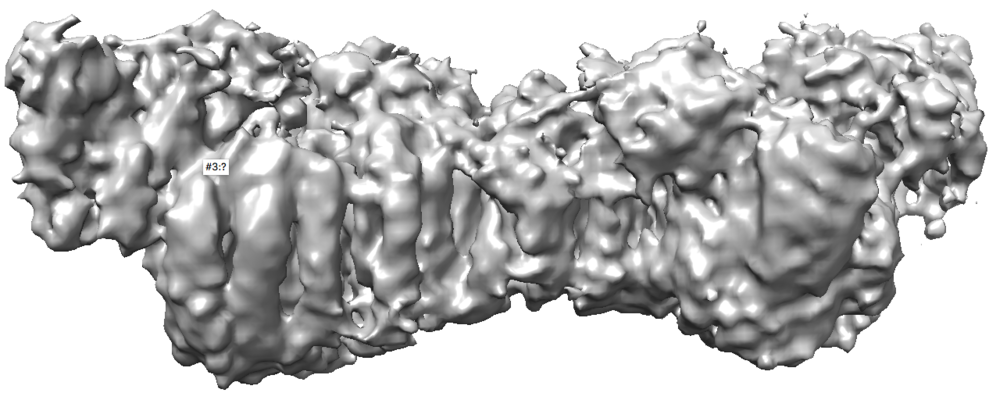
 Downloads & links
 emd_11837.png
 http://ftp.pdbj.org/pub/emdb/structures/EMD-11837


 Z (Sec.)
 Y (Row.)
 X (Col.)


 Sample components
 Processing
 Electron microscopy
 FIELD EMISSION GUN