 ムービー
ムービー コントローラー


+ データを開く

- 基本情報
基本情報
| 登録情報 | データベース: EMDB / ID: EMD-1086 | |||||||||
|---|---|---|---|---|---|---|---|---|---|---|
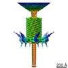
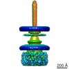
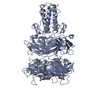
| タイトル | Three-dimensional rearrangement of proteins in the tail of bacteriophage T4 on infection of its host. | |||||||||
 マップデータ マップデータ | CryoEM density of the T4 tail with contracted sheath and the baseplate in the star conformation. | |||||||||
 試料 試料 |
| |||||||||
| 機能・相同性 |  機能・相同性情報 機能・相同性情報virus tail, sheath / symbiont genome ejection through host cell envelope, contractile tail mechanism / virus tail, baseplate / viral tail assembly / viral release from host cell / virion component / identical protein binding 類似検索 - 分子機能 | |||||||||
| 生物種 |  Enterobacteria phage T4 (ファージ) Enterobacteria phage T4 (ファージ) | |||||||||
| 手法 | 単粒子再構成法 / クライオ電子顕微鏡法 / 解像度: 16.0 Å | |||||||||
 データ登録者 データ登録者 | Leiman PG / Chipman PR / Kostyuchenko VA / Mesyanzhinov VV / Rossmann MG | |||||||||
 引用 引用 | ジャーナル: Cell / 年: 2004 タイトル: Three-dimensional rearrangement of proteins in the tail of bacteriophage T4 on infection of its host. 著者: Petr G Leiman / Paul R Chipman / Victor A Kostyuchenko / Vadim V Mesyanzhinov / Michael G Rossmann /  要旨: The contractile tail of bacteriophage T4 undergoes major structural transitions when the virus attaches to the host cell surface. The baseplate at the distal end of the tail changes from a hexagonal ...The contractile tail of bacteriophage T4 undergoes major structural transitions when the virus attaches to the host cell surface. The baseplate at the distal end of the tail changes from a hexagonal to a star shape. This causes the sheath around the tail tube to contract and the tail tube to protrude from the baseplate and pierce the outer cell membrane and the cell wall before reaching the inner cell membrane for subsequent viral DNA injection. Analogously, the T4 tail can be contracted by treatment with 3 M urea. The structure of the T4 contracted tail, including the head-tail joining region, has been determined by cryo-electron microscopy to 17 A resolution. This 1200 A-long, 20 MDa structure has been interpreted in terms of multiple copies of its approximately 20 component proteins. A comparison with the metastable hexagonal baseplate of the mature virus shows that the baseplate proteins move as rigid bodies relative to each other during the structural change. | |||||||||
| 履歴 |
|
- 構造の表示
構造の表示
| ムービー |
ムービービューア |
|---|---|
| 構造ビューア | EMマップ: SurfViewMolmilJmol/JSmol |
| 添付画像 |
- ダウンロードとリンク
-EMDBアーカイブ
| マップデータ | emd_1086.map.gz | 7.8 MB | EMDBマップデータ形式 | |
|---|---|---|---|---|
| ヘッダ (付随情報) | emd-1086-v30.xmlemd-1086.xml | 11.5 KB 11.5 KB | 表示 表示 | EMDBヘッダ |
| 画像 |  1086.gif 1086.gif | 11.9 KB | ||
| アーカイブディレクトリ |  http://ftp.pdbj.org/pub/emdb/structures/EMD-1086ftp://ftp.pdbj.org/pub/emdb/structures/EMD-1086 http://ftp.pdbj.org/pub/emdb/structures/EMD-1086ftp://ftp.pdbj.org/pub/emdb/structures/EMD-1086 | HTTPS FTP |
-検証レポート
| 文書・要旨 | emd_1086_validation.pdf.gz | 364.3 KB | 表示 | EMDB検証レポート |
|---|---|---|---|---|
| 文書・詳細版 | emd_1086_full_validation.pdf.gz | 363.9 KB | 表示 | |
| XML形式データ | emd_1086_validation.xml.gz | 6.5 KB | 表示 | |
| アーカイブディレクトリ | https://ftp.pdbj.org/pub/emdb/validation_reports/EMD-1086ftp://ftp.pdbj.org/pub/emdb/validation_reports/EMD-1086 | HTTPS FTP |
-関連構造データ
-リンク
| EMDBのページ | EMDB (EBI/PDBe) / EMDataResource |
|---|
-マップ
| ファイル | ダウンロード / ファイル: emd_1086.map.gz / 形式: CCP4 / 大きさ: 62.5 MB / タイプ: IMAGE STORED AS FLOATING POINT NUMBER (4 BYTES) | ||||||||||||||||||||||||||||||||||||||||||||||||||||||||||||||||||||
|---|---|---|---|---|---|---|---|---|---|---|---|---|---|---|---|---|---|---|---|---|---|---|---|---|---|---|---|---|---|---|---|---|---|---|---|---|---|---|---|---|---|---|---|---|---|---|---|---|---|---|---|---|---|---|---|---|---|---|---|---|---|---|---|---|---|---|---|---|---|
| 注釈 | CryoEM density of the T4 tail with contracted sheath and the baseplate in the star conformation. | ||||||||||||||||||||||||||||||||||||||||||||||||||||||||||||||||||||
| ボクセルのサイズ | X=Y=Z: 3.93285 Å | ||||||||||||||||||||||||||||||||||||||||||||||||||||||||||||||||||||
| 密度 |
| ||||||||||||||||||||||||||||||||||||||||||||||||||||||||||||||||||||
| 対称性 | 空間群: 1 | ||||||||||||||||||||||||||||||||||||||||||||||||||||||||||||||||||||
| 詳細 | EMDB XML:
CCP4マップ ヘッダ情報:
| ||||||||||||||||||||||||||||||||||||||||||||||||||||||||||||||||||||
-添付データ
- 試料の構成要素
-全体 : T4 phages treated with 3 M urea
| 全体 | 名称: T4 phages treated with 3 M urea |
|---|---|
| 要素 |
|
-超分子 #1000: T4 phages treated with 3 M urea
| 超分子 | 名称: T4 phages treated with 3 M urea / タイプ: sample / ID: 1000 / Number unique components: 1 |
|---|---|
| 分子量 | 実験値: 220 MDa / 理論値: 220 MDa / 手法: Estimate |
-超分子 #1: Enterobacteria phage T4
| 超分子 | 名称: Enterobacteria phage T4 / タイプ: virus / ID: 1 / Name.synonym: phage T4 / 詳細: treated with 3 M urea / NCBI-ID: 10665 / 生物種: Enterobacteria phage T4 / ウイルスタイプ: VIRION / ウイルス・単離状態: STRAIN / ウイルス・エンベロープ: No / ウイルス・中空状態: No / Syn species name: phage T4 |
|---|---|
| 宿主 | 生物種:  |
| 分子量 | 実験値: 220 MDa / 理論値: 220 MDa |
-実験情報
-構造解析
| 手法 | クライオ電子顕微鏡法 |
|---|---|
 解析 解析 | 単粒子再構成法 |
| 試料の集合状態 | particle |
-試料調製
| 濃度 | 5 mg/mL |
|---|---|
| 緩衝液 | pH: 7.5 / 詳細: H2O |
| グリッド | 詳細: 200 mesh cupper grid |
| 凍結 | 凍結剤: ETHANE / チャンバー内温度: 100 K |
- 電子顕微鏡法
| 顕微鏡 | FEI/PHILIPS CM300FEG/T |
|---|---|
| 温度 | 平均: 100 K |
| 特殊光学系 | エネルギーフィルター - 名称: FEI |
| 日付 | 2002年1月6日 |
| 撮影 | カテゴリ: FILM / フィルム・検出器のモデル: KODAK SO-163 FILM / デジタル化 - スキャナー: ZEISS SCAI / デジタル化 - サンプリング間隔: 3.93285 µm / 実像数: 100 / 平均電子線量: 20 e/Å2 / ビット/ピクセル: 8 |
| Tilt angle min | 0 |
| Tilt angle max | 0 |
| 電子線 | 加速電圧: 300 kV / 電子線源:  FIELD EMISSION GUN FIELD EMISSION GUN |
| 電子光学系 | 倍率(補正後): 47000 / 照射モード: SPOT SCAN / 撮影モード: BRIGHT FIELD / 最大 デフォーカス(公称値): 3.4 µm / 最小 デフォーカス(公称値): 0.5 µm / 倍率(公称値): 45000 |
| 試料ステージ | 試料ホルダー: Side entry liquid nitrogen-cooled cryo specimen holder 試料ホルダーモデル: GATAN LIQUID NITROGEN |
-画像解析
| CTF補正 | 詳細: Each particle |
|---|---|
| 最終 再構成 | 想定した対称性 - 点群: C6 (6回回転対称) / アルゴリズム: OTHER / 解像度のタイプ: BY AUTHOR / 解像度: 16.0 Å / 解像度の算出法: FSC 0.5 CUT-OFF / ソフトウェア - 名称: Spider / 使用した粒子像数: 1965 |
| 最終 角度割当 | 詳細: theta 45 degrees, phi 180 degrees |
-原子モデル構築 1
| 初期モデル | PDB ID: |
|---|---|
| ソフトウェア | 名称: Situs v.2.0 |
| 詳細 | The components were separately fitted using Situs v.2.0 |
| 精密化 | 空間: REAL |
| 得られたモデル | 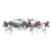 PDB-1tja: 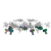 PDB-2fl9: 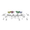 PDB-3foi: 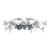 PDB-3h3y: 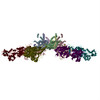 PDB-3j2n: |

-原子モデル構築 2
| 初期モデル | PDB ID: |
|---|---|
| ソフトウェア | 名称: Situs v.2.0 |
| 詳細 | The components were separately fitted using Situs v.2.0 |
| 精密化 | 空間: REAL |
| 得られたモデル | PDB-1tja: PDB-2fl9: PDB-3foi: PDB-3h3y: PDB-3j2n: |
-原子モデル構築 3
| 初期モデル | PDB ID: |
|---|---|
| ソフトウェア | 名称: Situs v.2.0 |
| 詳細 | The components were separately fitted using Situs v.2.0 |
| 精密化 | 空間: REAL |
| 得られたモデル | PDB-1tja: PDB-2fl9: PDB-3foi: PDB-3h3y: PDB-3j2n: |

