- EMDB-10502: Cryo-EM structure of p62-PB1 filament (L-type) -
+
Open data
ID or keywords:
Loading...
-
Basic information
Entry
Database: EMDB / ID: EMD-10502
Title
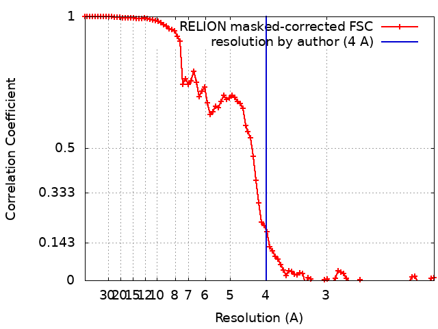
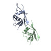
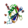
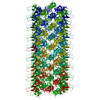
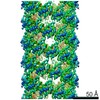
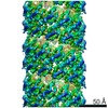
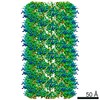
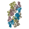
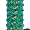
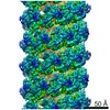
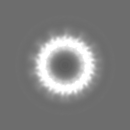
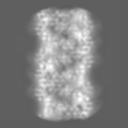
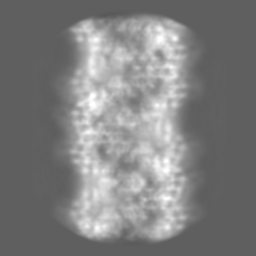
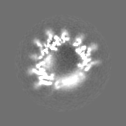
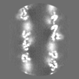
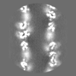
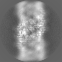
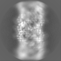
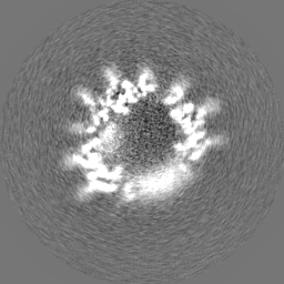
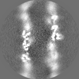
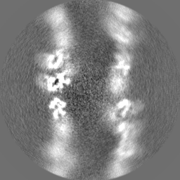
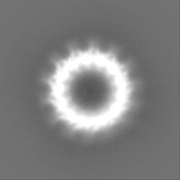
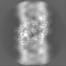
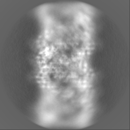
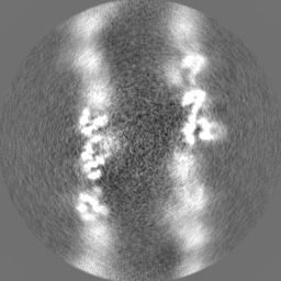
Cryo-EM structure of p62-PB1 filament (L-type)
Map data
Globally sharpened map
Sample
Complex: p62-PB1 domain filament (L-type)
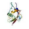
Protein or peptide: Sequestosome-1
Keywords
Autophagy / CYTOSOLIC PROTEIN
Function / homology
Function and homology information
protein localization to perinuclear region of cytoplasm / brown fat cell proliferation / protein targeting to vacuole involved in autophagy / regulation of Ras protein signal transduction / intracellular membraneless organelle / aggrephagy / negative regulation of toll-like receptor 4 signaling pathway / response to mitochondrial depolarisation / amphisome / regulation of protein complex stability ...protein localization to perinuclear region of cytoplasm / brown fat cell proliferation / protein targeting to vacuole involved in autophagy / regulation of Ras protein signal transduction / intracellular membraneless organelle / aggrephagy / negative regulation of toll-like receptor 4 signaling pathway / response to mitochondrial depolarisation / amphisome / regulation of protein complex stability / autophagy of mitochondrion / endosome organization / pexophagy / membraneless organelle assembly / regulation of mitochondrion organization / phagophore assembly site / ubiquitin-modified protein reader activity / regulation of canonical NF-kappaB signal transduction / Nuclear events mediated by NFE2L2 / aggresome / K63-linked polyubiquitin modification-dependent protein binding / endosomal transport / cellular response to stress / Lewy body / temperature homeostasis / negative regulation of ferroptosis / autolysosome / molecular sequestering activity / immune system process / mitophagy / energy homeostasis / inclusion body / signaling adaptor activity / negative regulation of protein ubiquitination / positive regulation of autophagy / ionotropic glutamate receptor binding / SH2 domain binding / autophagosome / p75NTR recruits signalling complexes / response to ischemia / NF-kB is activated and signals survival / Pexophagy / NRIF signals cell death from the nucleus / protein kinase C binding / protein catabolic process / positive regulation of long-term synaptic potentiation / positive regulation of protein localization to plasma membrane / PINK1-PRKN Mediated Mitophagy / sarcomere / macroautophagy / ubiquitin binding / protein sequestering activity / molecular condensate scaffold activity / P-body / receptor tyrosine kinase binding / PML body / autophagy / Interleukin-1 signaling / protein import into nucleus / Signaling by ALK fusions and activated point mutants / intracellular protein localization / late endosome / KEAP1-NFE2L2 pathway / signaling receptor activity / Neddylation / sperm midpiece / transcription by RNA polymerase II / ubiquitin-dependent protein catabolic process / protein-macromolecule adaptor activity / cell differentiation / intracellular signal transduction / positive regulation of apoptotic process / apoptotic process / ubiquitin protein ligase binding / protein kinase binding / protein-containing complex binding / glutamatergic synapse / enzyme binding / negative regulation of transcription by RNA polymerase II / endoplasmic reticulum / positive regulation of transcription by RNA polymerase II / mitochondrion / extracellular exosome / zinc ion binding / nucleoplasm / identical protein binding / cytoplasm / cytosol Similarity search - Function
Journal: Nat Commun / Year: 2020 Title: Structural basis of p62/SQSTM1 helical filaments and their role in cellular cargo uptake. Authors: Arjen J Jakobi / Stefan T Huber / Simon A Mortensen / Sebastian W Schultz / Anthimi Palara / Tanja Kuhm / Birendra Kumar Shrestha / Trond Lamark / Wim J H Hagen / Matthias Wilmanns / Terje ...Authors: Arjen J Jakobi / Stefan T Huber / Simon A Mortensen / Sebastian W Schultz / Anthimi Palara / Tanja Kuhm / Birendra Kumar Shrestha / Trond Lamark / Wim J H Hagen / Matthias Wilmanns / Terje Johansen / Andreas Brech / Carsten Sachse / Abstract: p62/SQSTM1 is an autophagy receptor and signaling adaptor with an N-terminal PB1 domain that forms the scaffold of phase-separated p62 bodies in the cell. The molecular determinants that govern PB1 ...p62/SQSTM1 is an autophagy receptor and signaling adaptor with an N-terminal PB1 domain that forms the scaffold of phase-separated p62 bodies in the cell. The molecular determinants that govern PB1 domain filament formation in vitro remain to be determined and the role of p62 filaments inside the cell is currently unclear. We here determine four high-resolution cryo-EM structures of different human and Arabidopsis PB1 domain assemblies and observed a filamentous ultrastructure of p62/SQSTM1 bodies using correlative cellular EM. We show that oligomerization or polymerization, driven by a double arginine finger in the PB1 domain, is a general requirement for lysosomal targeting of p62. Furthermore, the filamentous assembly state of p62 is required for autophagosomal processing of the p62-specific cargo KEAP1. Our results show that using such mechanisms, p62 filaments can be critical for cargo uptake in autophagy and are an integral part of phase-separated p62 bodies.
History
Deposition
Nov 18, 2019
-
Header (metadata) release
Jan 29, 2020
-
Map release
Feb 12, 2020
-
Update
May 22, 2024
-
Current status
May 22, 2024
Processing site: PDBe / Status: Released
-
Structure visualization
Movie
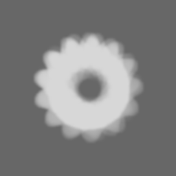
Surface view with section colored by density value
In the structure databanks used in Yorodumi, some data are registered as the other names, "COVID-19 virus" and "2019-nCoV". Here are the details of the virus and the list of structure data.
Jan 31, 2019. EMDB accession codes are about to change! (news from PDBe EMDB page)
EMDB accession codes are about to change! (news from PDBe EMDB page)
The allocation of 4 digits for EMDB accession codes will soon come to an end. Whilst these codes will remain in use, new EMDB accession codes will include an additional digit and will expand incrementally as the available range of codes is exhausted. The current 4-digit format prefixed with “EMD-” (i.e. EMD-XXXX) will advance to a 5-digit format (i.e. EMD-XXXXX), and so on. It is currently estimated that the 4-digit codes will be depleted around Spring 2019, at which point the 5-digit format will come into force.
The EM Navigator/Yorodumi systems omit the EMD- prefix.
Related info.:Q: What is EMD? / ID/Accession-code notation in Yorodumi/EM Navigator
Yorodumi is a browser for structure data from EMDB, PDB, SASBDB, etc.
This page is also the successor to EM Navigator detail page, and also detail information page/front-end page for Omokage search.
The word "yorodu" (or yorozu) is an old Japanese word meaning "ten thousand". "mi" (miru) is to see.
Related info.:EMDB / PDB / SASBDB / Comparison of 3 databanks / Yorodumi Search / Aug 31, 2016. New EM Navigator & Yorodumi / Yorodumi Papers / Jmol/JSmol / Function and homology information / Changes in new EM Navigator and Yorodumi
 Movie
Movie Controller


 Open data
 Basic information
 Map data
 Sample
 Keywords
 Function and homology information
 Homo sapiens (human)
 Authors
 Germany, 3 items
  Citation

 Structure visualization
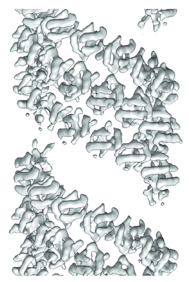
 Downloads & links
 emd_10502.png
 http://ftp.pdbj.org/pub/emdb/structures/EMD-10502
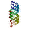


 Z (Sec.)
 Y (Row.)
 X (Col.)


 Sample components
 Processing
 Electron microscopy
 FIELD EMISSION GUN