Movie
Movie Controller


+ Open data

- Basic information
| Entry | Database: EMDB / ID: EMD-10081 | |||||||||
|---|---|---|---|---|---|---|---|---|---|---|
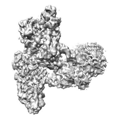
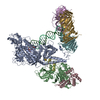
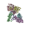
| Title | Human polymerase delta holoenzyme Conformer 2 | |||||||||
 Map data Map data | ||||||||||
 Sample Sample |
| |||||||||
 Keywords Keywords | Protein / REPLICATION | |||||||||
| Function / homology |  Function and homology information Function and homology informationdelta DNA polymerase complex / DNA synthesis involved in UV-damage excision repair / zeta DNA polymerase complex / nucleotide-excision repair complex / Cytosolic iron-sulfur cluster assembly / positive regulation of deoxyribonuclease activity / dinucleotide insertion or deletion binding / PCNA-p21 complex / mitotic telomere maintenance via semi-conservative replication / purine-specific mismatch base pair DNA N-glycosylase activity ...delta DNA polymerase complex / DNA synthesis involved in UV-damage excision repair / zeta DNA polymerase complex / nucleotide-excision repair complex / Cytosolic iron-sulfur cluster assembly / positive regulation of deoxyribonuclease activity / dinucleotide insertion or deletion binding / PCNA-p21 complex / mitotic telomere maintenance via semi-conservative replication / purine-specific mismatch base pair DNA N-glycosylase activity / 3'-5'-DNA exonuclease activity / nucleotide-excision repair, DNA gap filling / nuclear lamina / positive regulation of DNA-directed DNA polymerase activity / Polymerase switching / Telomere C-strand (Lagging Strand) Synthesis / MutLalpha complex binding / Processive synthesis on the lagging strand / DNA replication proofreading / PCNA complex / Removal of the Flap Intermediate / Processive synthesis on the C-strand of the telomere / Polymerase switching on the C-strand of the telomere / Removal of the Flap Intermediate from the C-strand / Mismatch repair (MMR) directed by MSH2:MSH3 (MutSbeta) / Mismatch repair (MMR) directed by MSH2:MSH6 (MutSalpha) / Transcription of E2F targets under negative control by DREAM complex / replisome / aggresome / Hydrolases; Acting on ester bonds; Exodeoxyribonucleases producing 5'-phosphomonoesters / error-free translesion synthesis / response to L-glutamate / DNA biosynthetic process / DNA synthesis involved in DNA repair / DNA strand elongation involved in DNA replication / histone acetyltransferase binding / DNA polymerase processivity factor activity / leading strand elongation / G1/S-Specific Transcription / response to dexamethasone / replication fork processing / nuclear replication fork / SUMOylation of DNA replication proteins / PCNA-Dependent Long Patch Base Excision Repair / fatty acid homeostasis / error-prone translesion synthesis / translesion synthesis / mismatch repair / response to cadmium ion / response to UV / estrous cycle / cyclin-dependent protein kinase holoenzyme complex / base-excision repair, gap-filling / DNA polymerase binding / positive regulation of endothelial cell proliferation / epithelial cell differentiation / positive regulation of DNA repair / male germ cell nucleus / TP53 Regulates Transcription of Genes Involved in G2 Cell Cycle Arrest / Translesion synthesis by REV1 / Translesion synthesis by POLK / liver regeneration / Translesion synthesis by POLI / replication fork / Gap-filling DNA repair synthesis and ligation in GG-NER / positive regulation of DNA replication / nuclear estrogen receptor binding / Termination of translesion DNA synthesis / Recognition of DNA damage by PCNA-containing replication complex / Translesion Synthesis by POLH / receptor tyrosine kinase binding / HDR through Homologous Recombination (HRR) / Dual Incision in GG-NER / cellular response to hydrogen peroxide / DNA-templated DNA replication / Dual incision in TC-NER / Gap-filling DNA repair synthesis and ligation in TC-NER / cellular response to UV / cellular response to xenobiotic stimulus / E3 ubiquitin ligases ubiquitinate target proteins / response to estradiol / heart development / 4 iron, 4 sulfur cluster binding / protein-macromolecule adaptor activity / damaged DNA binding / DNA-directed DNA polymerase / DNA-directed DNA polymerase activity / chromosome, telomeric region / DNA replication / nuclear body / DNA repair / nucleotide binding / centrosome / chromatin binding / protein-containing complex binding / chromatin / enzyme binding / negative regulation of transcription by RNA polymerase II / DNA binding / extracellular exosome Similarity search - Function | |||||||||
| Biological species |  Homo sapiens (human) / synthetic construct (others) Homo sapiens (human) / synthetic construct (others) | |||||||||
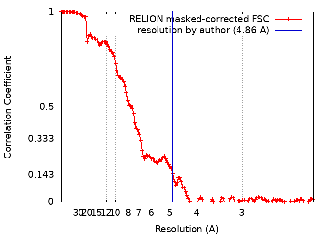
| Method | single particle reconstruction / cryo EM / Resolution: 4.86 Å | |||||||||
 Authors Authors | Lancey C / Hamdan SM / De Biasio A / Rashid F / Merino N / Ragan TJ / Savva C / Zaher MS / Blanco FJ | |||||||||
| Funding support |  United Kingdom, 1 items United Kingdom, 1 items
| |||||||||
 Citation Citation | Journal: Nat Commun / Year: 2020 Title: Structure of the processive human Pol δ holoenzyme. Authors: Claudia Lancey / Muhammad Tehseen / Vlad-Stefan Raducanu / Fahad Rashid / Nekane Merino / Timothy J Ragan / Christos G Savva / Manal S Zaher / Afnan Shirbini / Francisco J Blanco / Samir M ...Authors: Claudia Lancey / Muhammad Tehseen / Vlad-Stefan Raducanu / Fahad Rashid / Nekane Merino / Timothy J Ragan / Christos G Savva / Manal S Zaher / Afnan Shirbini / Francisco J Blanco / Samir M Hamdan / Alfredo De Biasio /   Abstract: In eukaryotes, DNA polymerase δ (Pol δ) bound to the proliferating cell nuclear antigen (PCNA) replicates the lagging strand and cooperates with flap endonuclease 1 (FEN1) to process the Okazaki ...In eukaryotes, DNA polymerase δ (Pol δ) bound to the proliferating cell nuclear antigen (PCNA) replicates the lagging strand and cooperates with flap endonuclease 1 (FEN1) to process the Okazaki fragments for their ligation. We present the high-resolution cryo-EM structure of the human processive Pol δ-DNA-PCNA complex in the absence and presence of FEN1. Pol δ is anchored to one of the three PCNA monomers through the C-terminal domain of the catalytic subunit. The catalytic core sits on top of PCNA in an open configuration while the regulatory subunits project laterally. This arrangement allows PCNA to thread and stabilize the DNA exiting the catalytic cleft and recruit FEN1 to one unoccupied monomer in a toolbelt fashion. Alternative holoenzyme conformations reveal important functional interactions that maintain PCNA orientation during synthesis. This work sheds light on the structural basis of Pol δ's activity in replicating the human genome. | |||||||||
| History |
|
- Structure visualization
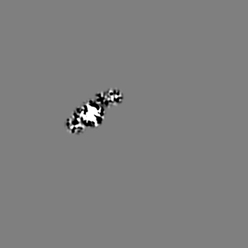
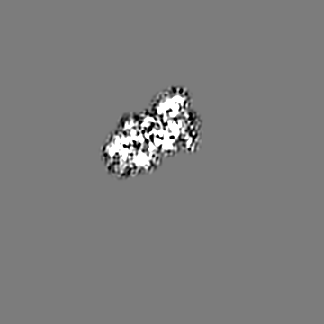
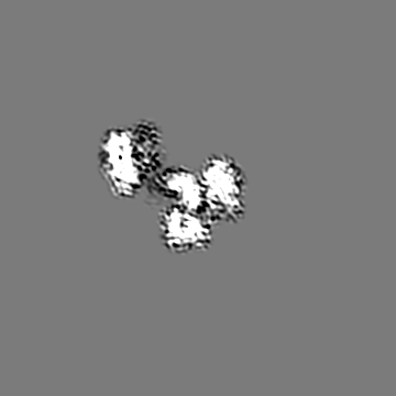
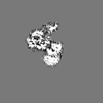
| Movie |
Movie viewer |
|---|---|
| Structure viewer | EM map: SurfViewMolmilJmol/JSmol |
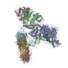
| Supplemental images |
- Downloads & links
-EMDB archive
| Map data | emd_10081.map.gz | 11 MB | EMDB map data format | |
|---|---|---|---|---|
| Header (meta data) | emd-10081-v30.xmlemd-10081.xml | 25.4 KB 25.4 KB | Display Display | EMDB header |
| FSC (resolution estimation) | emd_10081_fsc.xml | 12.9 KB | Display | FSC data file |
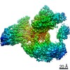
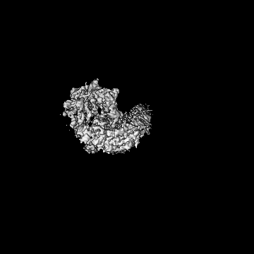
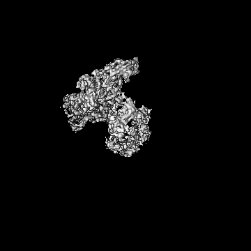
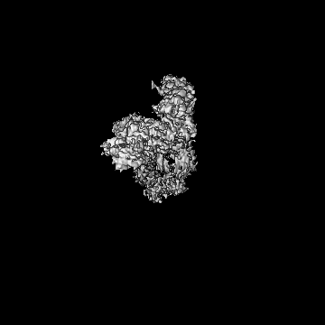
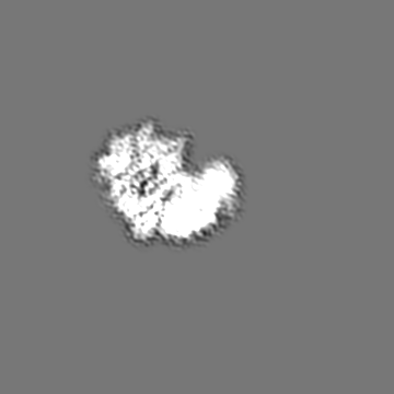
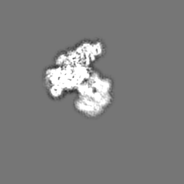
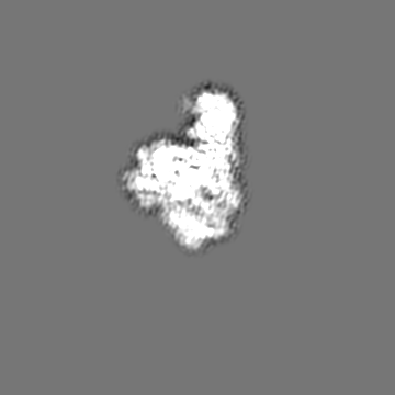
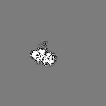
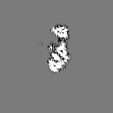
| Images |  emd_10081.png emd_10081.png | 62.8 KB | ||
| Filedesc metadata | emd-10081.cif.gz | 8.4 KB | ||
| Archive directory |  http://ftp.pdbj.org/pub/emdb/structures/EMD-10081ftp://ftp.pdbj.org/pub/emdb/structures/EMD-10081 http://ftp.pdbj.org/pub/emdb/structures/EMD-10081ftp://ftp.pdbj.org/pub/emdb/structures/EMD-10081 | HTTPS FTP |
-Related structure data
| Related structure data |  6s1nMC  6s1mC  6s1oC  6tnyC  6tnzC C: citing same article ( M: atomic model generated by this map |
|---|---|
| Similar structure data | |
| EM raw data | EMPIAR-10824 (Title: Cryo electron micrographs of Pol delta-DNA-PCNA giving rise to various PCNA tilt angles Data size: 3.6 TB Data #1: Cryo-electron micrographs of Pol delta holoenzyme giving rise to different tilted conformers, collection 2 [micrographs - multiframe] Data #2: Cryo-electron micrographs of Pol delta holoenzyme giving rise to different tilted conformers, collection 1 [micrographs - multiframe] Data #3: Cryo-electron micrographs of Pol delta holoenzyme giving rise to different tilted conformers, collection 3 [micrographs - multiframe]) |


-Links
| EMDB pages | EMDB (EBI/PDBe) / EMDataResource |
|---|---|
| Related items in Molecule of the Month |


-Map
| File | Download / File: emd_10081.map.gz / Format: CCP4 / Size: 178 MB / Type: IMAGE STORED AS FLOATING POINT NUMBER (4 BYTES) | ||||||||||||||||||||||||||||||||||||||||||||||||||||||||||||||||||||
|---|---|---|---|---|---|---|---|---|---|---|---|---|---|---|---|---|---|---|---|---|---|---|---|---|---|---|---|---|---|---|---|---|---|---|---|---|---|---|---|---|---|---|---|---|---|---|---|---|---|---|---|---|---|---|---|---|---|---|---|---|---|---|---|---|---|---|---|---|---|
| Projections & slices | Image control
Images are generated by Spider. | ||||||||||||||||||||||||||||||||||||||||||||||||||||||||||||||||||||
| Voxel size | X=Y=Z: 1.08 Å | ||||||||||||||||||||||||||||||||||||||||||||||||||||||||||||||||||||
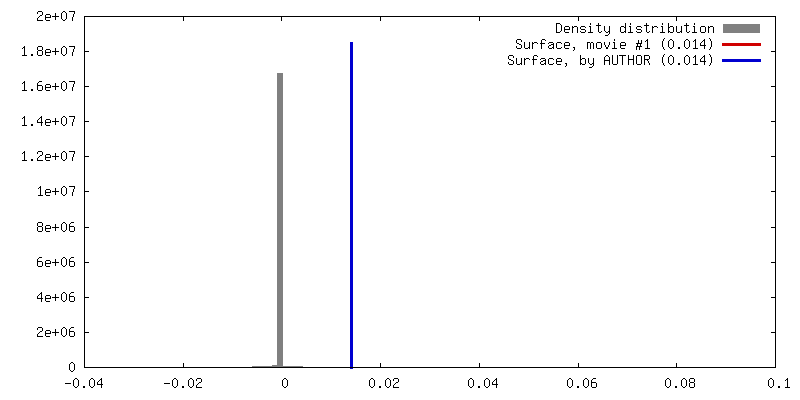
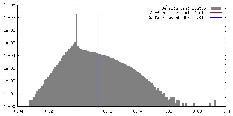
| Density |
| ||||||||||||||||||||||||||||||||||||||||||||||||||||||||||||||||||||
| Symmetry | Space group: 1 | ||||||||||||||||||||||||||||||||||||||||||||||||||||||||||||||||||||
| Details | EMDB XML:
CCP4 map header:
| ||||||||||||||||||||||||||||||||||||||||||||||||||||||||||||||||||||
 Z (Sec.)
 Y (Row.)
 X (Col.)


-Supplemental data
- Sample components
+Entire : PolD holoenzyme
+Supramolecule #1: PolD holoenzyme
+Supramolecule #2: Human polymerase delta
+Supramolecule #3: Proliferating cell nuclear antigen
+Supramolecule #4: DNA
+Macromolecule #1: DNA polymerase delta catalytic subunit

 Spodoptera frugiperda (fall armyworm)
+Macromolecule #2: DNA polymerase delta subunit 2
+Macromolecule #3: DNA polymerase delta subunit 3
+Macromolecule #4: DNA polymerase delta subunit 4
+Macromolecule #5: Proliferating cell nuclear antigen

+Macromolecule #6: DNA primer
+Macromolecule #7: DNA template
+Macromolecule #8: ZINC ION
+Macromolecule #9: IRON/SULFUR CLUSTER

+Macromolecule #10: THYMIDINE-5'-TRIPHOSPHATE

-Experimental details
-Structure determination
| Method | cryo EM |
|---|---|
 Processing Processing | single particle reconstruction |
| Aggregation state | particle |
-Sample preparation
| Concentration | 0.355 mg/mL | |||||||||||||||
|---|---|---|---|---|---|---|---|---|---|---|---|---|---|---|---|---|
| Buffer | pH: 7.5 Component:
| |||||||||||||||
| Grid | Model: Quantifoil, UltrAuFoil, R1.2/1.3 / Material: GOLD / Mesh: 300 / Support film - Material: GRAPHENE OXIDE / Support film - topology: CONTINUOUS / Pretreatment - Type: GLOW DISCHARGE / Pretreatment - Time: 300 sec. | |||||||||||||||
| Vitrification | Cryogen name: ETHANE / Chamber humidity: 100 % / Chamber temperature: 277 K / Instrument: FEI VITROBOT MARK IV |
- Electron microscopy
| Microscope | FEI TITAN KRIOS |
|---|---|
| Temperature | Min: 77.0 K / Max: 77.0 K |
| Specialist optics | Phase plate: VOLTA PHASE PLATE |
| Image recording | Film or detector model: FEI FALCON III (4k x 4k) / Detector mode: COUNTING / Digitization - Dimensions - Width: 4096 pixel / Digitization - Dimensions - Height: 4096 pixel / Number grids imaged: 2 / Number real images: 3575 / Average exposure time: 60.0 sec. / Average electron dose: 35.0 e/Å2 |
| Electron beam | Acceleration voltage: 300 kV / Electron source:  FIELD EMISSION GUN FIELD EMISSION GUN |
| Electron optics | C2 aperture diameter: 50.0 µm / Calibrated magnification: 130000 / Illumination mode: FLOOD BEAM / Imaging mode: BRIGHT FIELD / Cs: 2.7 mm / Nominal defocus max: 0.6 µm / Nominal defocus min: 0.4 µm / Nominal magnification: 75000 |
| Sample stage | Specimen holder model: FEI TITAN KRIOS AUTOGRID HOLDER / Cooling holder cryogen: NITROGEN |
| Experimental equipment |  Model: Titan Krios / Image courtesy: FEI Company |