Movie
Movie Controller


+ Open data

- Basic information
| Entry | Database: EMDB / ID: EMD-10059 | |||||||||||||||
|---|---|---|---|---|---|---|---|---|---|---|---|---|---|---|---|---|
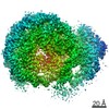
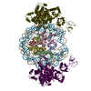
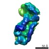
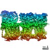
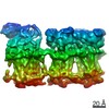
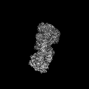
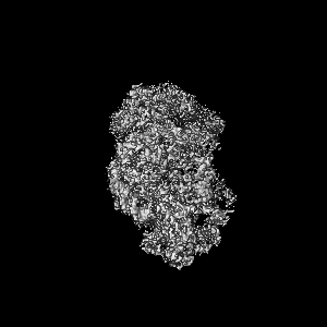
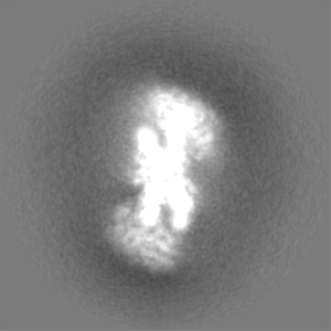
| Title | Nucleosome-CHD4 complex structure (two CHD4 copies) | |||||||||||||||
 Map data Map data | ||||||||||||||||
 Sample Sample |
| |||||||||||||||
 Keywords Keywords | Complex / ATPase / chromatin / nucleosome / CHD family / TRANSCRIPTION | |||||||||||||||
| Function / homology |  Function and homology information Function and homology informationcerebellar granule cell to Purkinje cell synapse / terminal button organization / NuRD complex / regulation of cell fate specification / NGF-stimulated transcription / regulation of stem cell differentiation / ATP-dependent chromatin remodeler activity / regulation of synapse assembly / RNA Polymerase I Transcription Initiation / Transcriptional regulation of brown and beige adipocyte differentiation by EBF2 ...cerebellar granule cell to Purkinje cell synapse / terminal button organization / NuRD complex / regulation of cell fate specification / NGF-stimulated transcription / regulation of stem cell differentiation / ATP-dependent chromatin remodeler activity / regulation of synapse assembly / RNA Polymerase I Transcription Initiation / Transcriptional regulation of brown and beige adipocyte differentiation by EBF2 / Regulation of TP53 Activity through Acetylation / site of DNA damage / Regulation of PTEN gene transcription / transcription coregulator binding / ERCC6 (CSB) and EHMT2 (G9a) positively regulate rRNA expression / Regulation of endogenous retroelements by KRAB-ZFP proteins / HDACs deacetylate histones / Regulation of endogenous retroelements by Piwi-interacting RNAs (piRNAs) / double-strand break repair via homologous recombination / RNA polymerase II transcription regulator complex / Hydrolases; Acting on acid anhydrides; Acting on acid anhydrides to facilitate cellular and subcellular movement / histone deacetylase binding / structural constituent of chromatin / transcription corepressor activity / nucleosome / nucleosome assembly / heterochromatin formation / histone binding / Potential therapeutics for SARS / RNA polymerase II-specific DNA-binding transcription factor binding / chromosome, telomeric region / chromatin remodeling / protein heterodimerization activity / negative regulation of gene expression / negative regulation of DNA-templated transcription / chromatin binding / centrosome / positive regulation of DNA-templated transcription / chromatin / negative regulation of transcription by RNA polymerase II / ATP hydrolysis activity / protein-containing complex / DNA binding / zinc ion binding / nucleoplasm / ATP binding / membrane / nucleus / cytoplasm Similarity search - Function | |||||||||||||||
| Biological species |  Homo sapiens (human) Homo sapiens (human) | |||||||||||||||
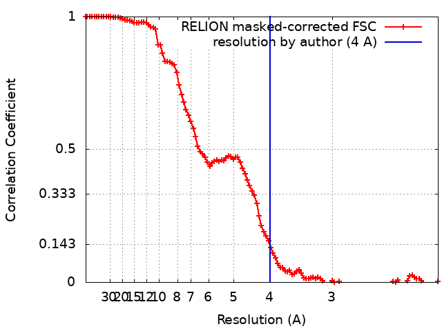
| Method | single particle reconstruction / cryo EM / Resolution: 4.0 Å | |||||||||||||||
 Authors Authors | Farnung L / Ochmann M | |||||||||||||||
| Funding support |  Germany, 4 items Germany, 4 items
| |||||||||||||||
 Citation Citation | Journal: Elife / Year: 2020 Title: Nucleosome-CHD4 chromatin remodeler structure maps human disease mutations. Authors: Lucas Farnung / Moritz Ochmann / Patrick Cramer / Abstract: Chromatin remodeling plays important roles in gene regulation during development, differentiation and in disease. The chromatin remodeling enzyme CHD4 is a component of the NuRD and ChAHP complexes ...Chromatin remodeling plays important roles in gene regulation during development, differentiation and in disease. The chromatin remodeling enzyme CHD4 is a component of the NuRD and ChAHP complexes that are involved in gene repression. Here, we report the cryo-electron microscopy (cryo-EM) structure of CHD4 engaged with a nucleosome core particle in the presence of the non-hydrolysable ATP analogue AMP-PNP at an overall resolution of 3.1 Å. The ATPase motor of CHD4 binds and distorts nucleosomal DNA at superhelical location (SHL) +2, supporting the 'twist defect' model of chromatin remodeling. CHD4 does not induce unwrapping of terminal DNA, in contrast to its homologue Chd1, which functions in gene activation. Our structure also maps CHD4 mutations that are associated with human cancer or the intellectual disability disorder Sifrim-Hitz-Weiss syndrome. | |||||||||||||||
| History |
|
- Structure visualization
| Movie |
Movie viewer |
|---|---|
| Structure viewer | EM map: SurfViewMolmilJmol/JSmol |
| Supplemental images |

- Downloads & links
-EMDB archive
| Map data | emd_10059.map.gz | 7.6 MB | EMDB map data format | |
|---|---|---|---|---|
| Header (meta data) | emd-10059-v30.xmlemd-10059.xml | 30.9 KB 30.9 KB | Display Display | EMDB header |
| FSC (resolution estimation) | emd_10059_fsc.xml | 10.7 KB | Display | FSC data file |
| Images | 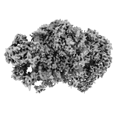 emd_10059.png emd_10059.png | 67.5 KB | ||
| Masks | emd_10059_msk_1.map | 103 MB | Mask map | |
| Filedesc metadata | emd-10059.cif.gz | 7.9 KB | ||
| Others | emd_10059_additional_1.map.gzemd_10059_additional_2.map.gzemd_10059_half_map_1.map.gzemd_10059_half_map_2.map.gz | 80.4 MB 95.9 MB 80.8 MB 80.8 MB | ||
| Archive directory |  http://ftp.pdbj.org/pub/emdb/structures/EMD-10059ftp://ftp.pdbj.org/pub/emdb/structures/EMD-10059 http://ftp.pdbj.org/pub/emdb/structures/EMD-10059ftp://ftp.pdbj.org/pub/emdb/structures/EMD-10059 | HTTPS FTP |
-Related structure data
| Related structure data | 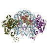 6ryuMC  6ryrC C: citing same article ( M: atomic model generated by this map |
|---|---|
| Similar structure data | |
| EM raw data | EMPIAR-10411 (Title: Single Particle Cryo-EM Reconstructions of NCP-CHD4 complexes Data size: 721.2 Data #1: Unaligned multi-frame micrographs of NCP-CHD4 Dataset 1 [micrographs - multiframe] Data #2: Averaged and aligned micrographs Dataset 1 [micrographs - single frame] Data #3: Unaligned multi-frame micrographs of NCP-CHD4 Dataset 2 [micrographs - multiframe] Data #4: Averaged and aligned micrographs NCP-CHD4 Dataset 2 [micrographs - single frame] Data #5: Unaligned multi-frame micrographs of NCP-CHD4 Dataset 3 [micrographs - multiframe] Data #6: Averaged and aligned micrographs Dataset 3 [micrographs - single frame]) |

-Links
| EMDB pages | EMDB (EBI/PDBe) / EMDataResource |
|---|---|
| Related items in Molecule of the Month |


-Map
| File | Download / File: emd_10059.map.gz / Format: CCP4 / Size: 103 MB / Type: IMAGE STORED AS FLOATING POINT NUMBER (4 BYTES) | ||||||||||||||||||||||||||||||||||||||||||||||||||||||||||||
|---|---|---|---|---|---|---|---|---|---|---|---|---|---|---|---|---|---|---|---|---|---|---|---|---|---|---|---|---|---|---|---|---|---|---|---|---|---|---|---|---|---|---|---|---|---|---|---|---|---|---|---|---|---|---|---|---|---|---|---|---|---|
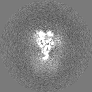
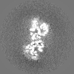
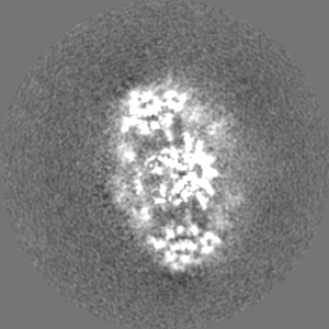
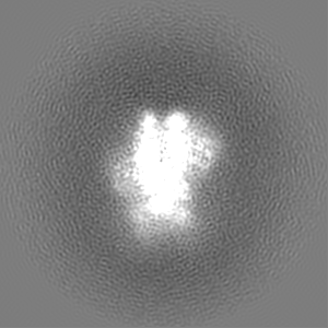
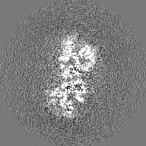
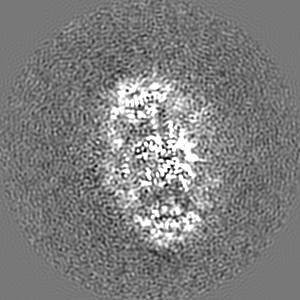
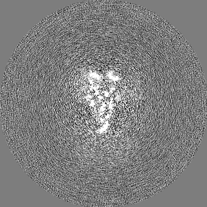
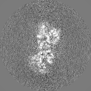
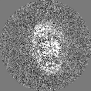
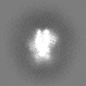
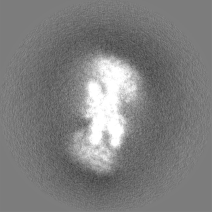
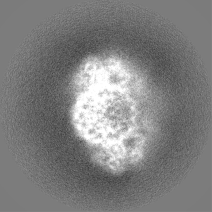
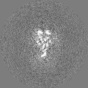
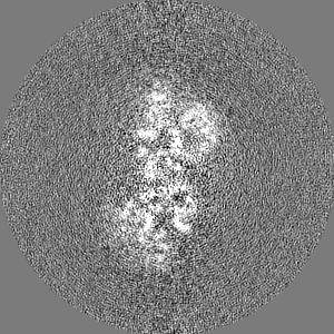
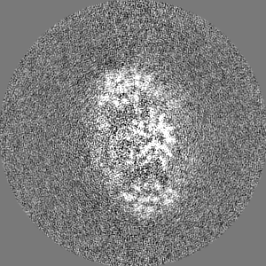
| Projections & slices | Image control
Images are generated by Spider. | ||||||||||||||||||||||||||||||||||||||||||||||||||||||||||||
| Voxel size | X=Y=Z: 1.05 Å | ||||||||||||||||||||||||||||||||||||||||||||||||||||||||||||
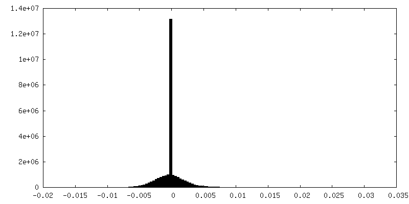
| Density |
| ||||||||||||||||||||||||||||||||||||||||||||||||||||||||||||
| Symmetry | Space group: 1 | ||||||||||||||||||||||||||||||||||||||||||||||||||||||||||||
| Details | EMDB XML:
CCP4 map header:
| ||||||||||||||||||||||||||||||||||||||||||||||||||||||||||||
 Z (Sec.)
 Y (Row.)
 X (Col.)


-Supplemental data
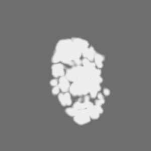
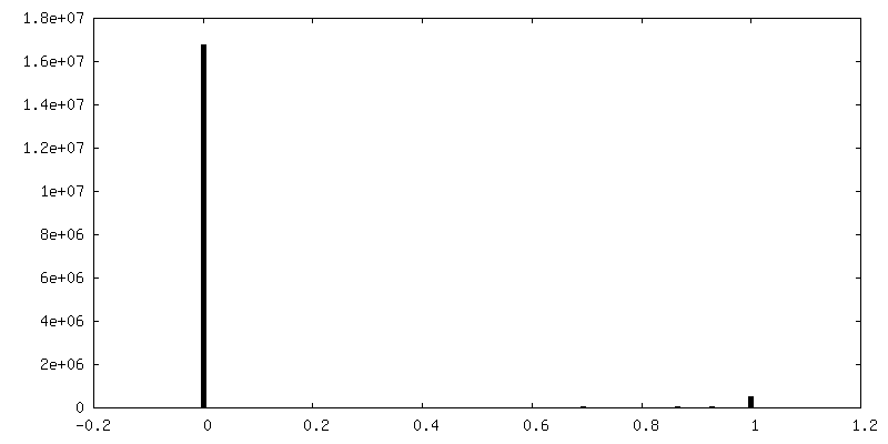
-Mask #1
| File | emd_10059_msk_1.map | ||||||||||||
|---|---|---|---|---|---|---|---|---|---|---|---|---|---|
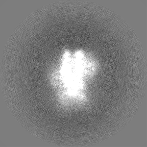
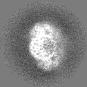
| Projections & Slices |
| ||||||||||||
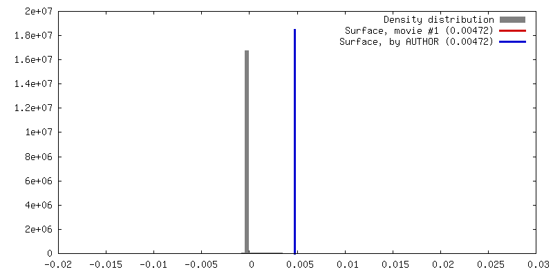
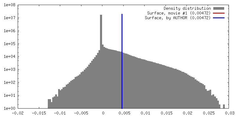
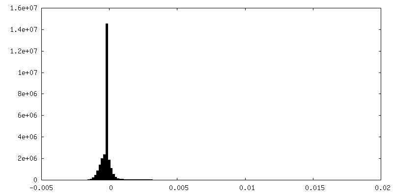
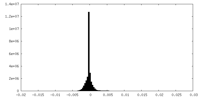
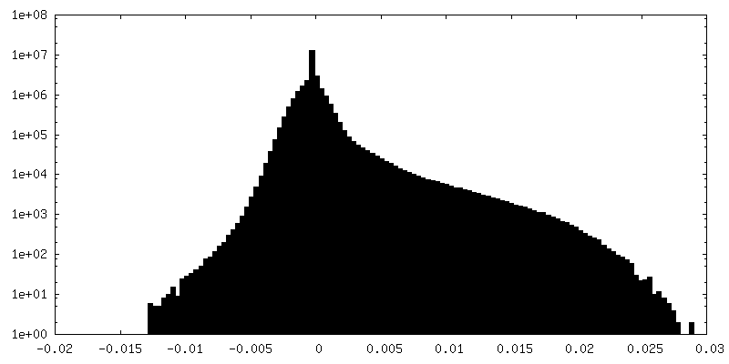
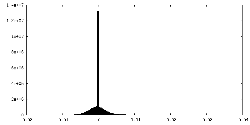
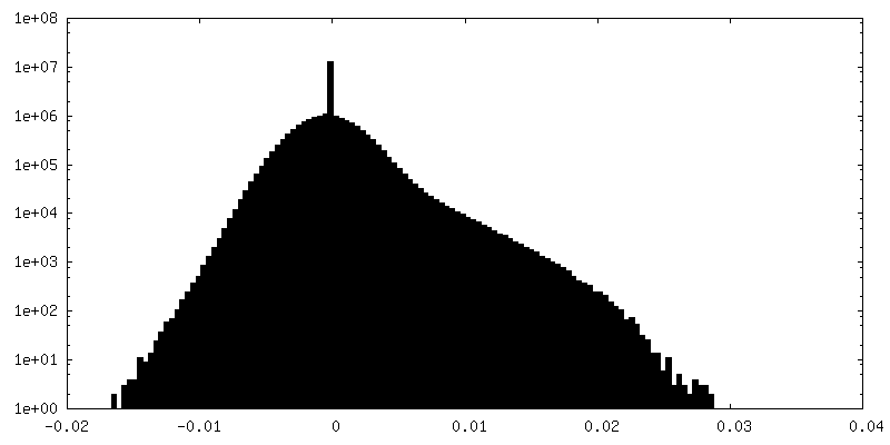
| Density Histograms |

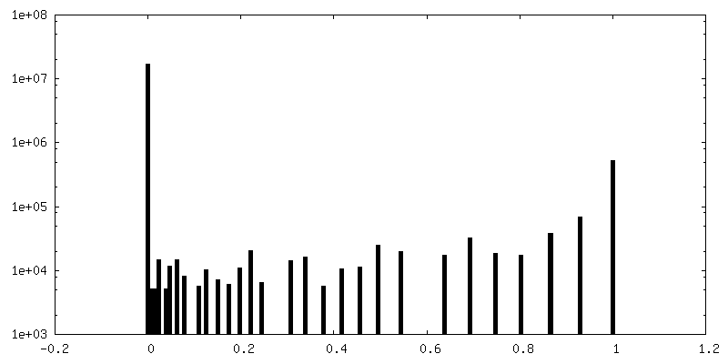
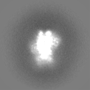
-Additional map: #1
| File | emd_10059_additional_1.map | ||||||||||||
|---|---|---|---|---|---|---|---|---|---|---|---|---|---|
| Projections & Slices |
| ||||||||||||
| Density Histograms |


-Additional map: #2
| File | emd_10059_additional_2.map | ||||||||||||
|---|---|---|---|---|---|---|---|---|---|---|---|---|---|
| Projections & Slices |
| ||||||||||||
| Density Histograms |


-Half map: #2
| File | emd_10059_half_map_1.map | ||||||||||||
|---|---|---|---|---|---|---|---|---|---|---|---|---|---|
| Projections & Slices |
| ||||||||||||
| Density Histograms |

-Half map: #1
| File | emd_10059_half_map_2.map | ||||||||||||
|---|---|---|---|---|---|---|---|---|---|---|---|---|---|
| Projections & Slices |
| ||||||||||||
| Density Histograms |
- Sample components
+Entire : Nucleosome-CHD4 complex
+Supramolecule #1: Nucleosome-CHD4 complex
+Supramolecule #2: Histone
+Supramolecule #3: DNA
+Supramolecule #4: Chromodomain-helicase-DNA-binding protein 4
+Macromolecule #1: Histone H3.2

+Macromolecule #2: Histone H4
+Macromolecule #3: Histone H2A type 1
+Macromolecule #4: Histone H2B 1.1
+Macromolecule #7: Chromodomain-helicase-DNA-binding protein 4,CHD4,Chromodomain-hel...
 Trichoplusia ni (cabbage looper)
+Macromolecule #5: DNA (149-MER)
+Macromolecule #6: DNA (149-MER)
+Macromolecule #8: PHOSPHOAMINOPHOSPHONIC ACID-ADENYLATE ESTER

+Macromolecule #9: MAGNESIUM ION
+Macromolecule #10: ZINC ION
-Experimental details
-Structure determination
| Method | cryo EM |
|---|---|
 Processing Processing | single particle reconstruction |
| Aggregation state | particle |
-Sample preparation
| Buffer | pH: 7.4 |
|---|---|
| Vitrification | Cryogen name: ETHANE / Chamber humidity: 100 % / Chamber temperature: 277 K / Instrument: FEI VITROBOT MARK IV |
- Electron microscopy
| Microscope | FEI TITAN KRIOS |
|---|---|
| Image recording | Film or detector model: GATAN K2 SUMMIT (4k x 4k) / Detector mode: COUNTING / Average electron dose: 43.0 e/Å2 |
| Electron beam | Acceleration voltage: 300 kV / Electron source:  FIELD EMISSION GUN FIELD EMISSION GUN |
| Electron optics | Illumination mode: SPOT SCAN / Imaging mode: BRIGHT FIELD |
| Sample stage | Specimen holder model: FEI TITAN KRIOS AUTOGRID HOLDER / Cooling holder cryogen: NITROGEN |
| Experimental equipment |  Model: Titan Krios / Image courtesy: FEI Company |