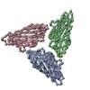
+ Open data
- Basic information
| Entry | Database: EMDB / ID: EMD-10003 | |||||||||
|---|---|---|---|---|---|---|---|---|---|---|
| Title | CryoEM structure of wild-type Turnip Yellows Virus | |||||||||
 Map data Map data | sharpened map | |||||||||
 Sample Sample |
| |||||||||
| Function / homology | Potato leaf roll virus readthrough protein / Potato leaf roll virus readthrough protein / Luteovirus group 1 coat protein / Luteovirus coat protein / host cell plasmodesma / host cell periplasmic space / viral capsid / structural molecule activity / Readthrough protein P3-RTD Function and homology information Function and homology information | |||||||||
| Biological species |  Beet western yellows virus-FL1 Beet western yellows virus-FL1 | |||||||||
| Method | single particle reconstruction / cryo EM / Resolution: 4.08 Å | |||||||||
 Authors Authors | Trapani S / Lai Kee Him J / Hoh F / Brault V / Bron P | |||||||||
| Funding support |  France, 1 items France, 1 items
| |||||||||
 Citation Citation |  Journal: To Be Published Journal: To Be PublishedTitle: CryoEM structure of Turnip Yellows Virus Authors: Trapani S / Lai Kee Him J / Boissinot S / Reinbold C / Fallet C / Ancelin A / Lecorre F / Hoh F / Ziegler-Graff V / Brault V / Bron P | |||||||||
| History |
|
- Structure visualization
| Movie |
 Movie viewer Movie viewer |
|---|---|
| Structure viewer | EM map:  SurfView SurfView Molmil Molmil Jmol/JSmol Jmol/JSmol |
| Supplemental images |
- Downloads & links
-EMDB archive
| Map data |  emd_10003.map.gz emd_10003.map.gz | 196.2 MB |  EMDB map data format EMDB map data format | |
|---|---|---|---|---|
| Header (meta data) |  emd-10003-v30.xml emd-10003-v30.xml emd-10003.xml emd-10003.xml | 17.1 KB 17.1 KB | Display Display |  EMDB header EMDB header |
| FSC (resolution estimation) |  emd_10003_fsc.xml emd_10003_fsc.xml | 13.5 KB | Display |  FSC data file FSC data file |
| Images |  emd_10003.png emd_10003.png | 247.5 KB | ||
| Others |  emd_10003_additional.map.gz emd_10003_additional.map.gz emd_10003_additional_1.map.gz emd_10003_additional_1.map.gz emd_10003_half_map_1.map.gz emd_10003_half_map_1.map.gz emd_10003_half_map_2.map.gz emd_10003_half_map_2.map.gz | 163.3 MB 163.3 MB 165.8 MB 165.8 MB | ||
| Archive directory |  http://ftp.pdbj.org/pub/emdb/structures/EMD-10003 http://ftp.pdbj.org/pub/emdb/structures/EMD-10003 ftp://ftp.pdbj.org/pub/emdb/structures/EMD-10003 ftp://ftp.pdbj.org/pub/emdb/structures/EMD-10003 | HTTPS FTP |
-Validation report
| Summary document |  emd_10003_validation.pdf.gz emd_10003_validation.pdf.gz | 394.9 KB | Display |  EMDB validaton report EMDB validaton report |
|---|---|---|---|---|
| Full document |  emd_10003_full_validation.pdf.gz emd_10003_full_validation.pdf.gz | 394 KB | Display | |
| Data in XML |  emd_10003_validation.xml.gz emd_10003_validation.xml.gz | 19.3 KB | Display | |
| Arichive directory |  https://ftp.pdbj.org/pub/emdb/validation_reports/EMD-10003 https://ftp.pdbj.org/pub/emdb/validation_reports/EMD-10003 ftp://ftp.pdbj.org/pub/emdb/validation_reports/EMD-10003 ftp://ftp.pdbj.org/pub/emdb/validation_reports/EMD-10003 | HTTPS FTP |
-Related structure data
| Related structure data |  9fhpM M: atomic model generated by this map |
|---|---|
| Similar structure data |
- Links
Links
| EMDB pages |  EMDB (EBI/PDBe) / EMDB (EBI/PDBe) /  EMDataResource EMDataResource |
|---|
- Map
Map
| File |  Download / File: emd_10003.map.gz / Format: CCP4 / Size: 209.3 MB / Type: IMAGE STORED AS FLOATING POINT NUMBER (4 BYTES) Download / File: emd_10003.map.gz / Format: CCP4 / Size: 209.3 MB / Type: IMAGE STORED AS FLOATING POINT NUMBER (4 BYTES) | ||||||||||||||||||||||||||||||||||||||||||||||||||||||||||||
|---|---|---|---|---|---|---|---|---|---|---|---|---|---|---|---|---|---|---|---|---|---|---|---|---|---|---|---|---|---|---|---|---|---|---|---|---|---|---|---|---|---|---|---|---|---|---|---|---|---|---|---|---|---|---|---|---|---|---|---|---|---|
| Annotation | sharpened map | ||||||||||||||||||||||||||||||||||||||||||||||||||||||||||||
| Projections & slices | Image control
Images are generated by Spider. | ||||||||||||||||||||||||||||||||||||||||||||||||||||||||||||
| Voxel size | X=Y=Z: 1.225 Å | ||||||||||||||||||||||||||||||||||||||||||||||||||||||||||||
| Density |
| ||||||||||||||||||||||||||||||||||||||||||||||||||||||||||||
| Symmetry | Space group: 1 | ||||||||||||||||||||||||||||||||||||||||||||||||||||||||||||
| Details | EMDB XML:
CCP4 map header:
| ||||||||||||||||||||||||||||||||||||||||||||||||||||||||||||
-Supplemental data
-Additional map: unsharpened map
| File | emd_10003_additional.map | ||||||||||||
|---|---|---|---|---|---|---|---|---|---|---|---|---|---|
| Annotation | unsharpened map | ||||||||||||
| Projections & Slices |
| ||||||||||||
| Density Histograms |
-Additional map: unsharpened map
| File | emd_10003_additional_1.map | ||||||||||||
|---|---|---|---|---|---|---|---|---|---|---|---|---|---|
| Annotation | unsharpened map | ||||||||||||
| Projections & Slices |
| ||||||||||||
| Density Histograms |
-Half map: half 1 map
| File | emd_10003_half_map_1.map | ||||||||||||
|---|---|---|---|---|---|---|---|---|---|---|---|---|---|
| Annotation | half_1 map | ||||||||||||
| Projections & Slices |
| ||||||||||||
| Density Histograms |
-Half map: half 2 map
| File | emd_10003_half_map_2.map | ||||||||||||
|---|---|---|---|---|---|---|---|---|---|---|---|---|---|
| Annotation | half_2 map | ||||||||||||
| Projections & Slices |
| ||||||||||||
| Density Histograms |
- Sample components
-Entire : Beet western yellows virus-FL1
| Entire | Name:  Beet western yellows virus-FL1 Beet western yellows virus-FL1 |
|---|---|
| Components |
|
-Supramolecule #1: Beet western yellows virus-FL1
| Supramolecule | Name: Beet western yellows virus-FL1 / type: virus / ID: 1 / Parent: 0 / Macromolecule list: all / NCBI-ID: 12043 / Sci species name: Beet western yellows virus-FL1 / Virus type: VIRION / Virus isolate: OTHER / Virus enveloped: No / Virus empty: No |
|---|---|
| Virus shell | Shell ID: 1 / Name: capsid / T number (triangulation number): 3 |
-Macromolecule #1: Major coat protein
| Macromolecule | Name: Major coat protein / type: protein_or_peptide / ID: 1 / Enantiomer: LEVO |
|---|---|
| Source (natural) | Organism:  Beet western yellows virus-FL1 Beet western yellows virus-FL1 |
| Sequence | String: MNTVVGRRII NGRRRPRRQT RRAQRPQPVV VVQTSRATQR RPRRRRRGNN RTGRTVPTRG AGSSETFVF SKDNLAGSSS GAITFGPSLS DCPAFSNGML KAYHEYKISM VILEFVSEAS S QNSGSIAY ELDPHCKLNS LSSTINKFGI TKPGKRAFTA SYINGTEWHD ...String: MNTVVGRRII NGRRRPRRQT RRAQRPQPVV VVQTSRATQR RPRRRRRGNN RTGRTVPTRG AGSSETFVF SKDNLAGSSS GAITFGPSLS DCPAFSNGML KAYHEYKISM VILEFVSEAS S QNSGSIAY ELDPHCKLNS LSSTINKFGI TKPGKRAFTA SYINGTEWHD VAEDQFRILY KG NGSSSIA GSFRITIKCQ FHNPK |
-Macromolecule #2: Minor capsid protein P3-RTD
| Macromolecule | Name: Minor capsid protein P3-RTD / type: protein_or_peptide / ID: 2 / Enantiomer: LEVO |
|---|---|
| Source (natural) | Organism:  Beet western yellows virus-FL1 Beet western yellows virus-FL1 |
| Sequence | String: MNTVVGRRII NGRRRPRRQT RRAQRPQPVV VVQTSRATQR RPRRRRRGNN RTGRTVPTRG AGSSETFVF SKDNLAGSSS GAITFGPSLS DCPAFSNGML KAYHEYKISM VILEFVSEAS S QNSGSIAY ELDPHCKLNS LSSTINKFGI TKPGKRAFTA SYINGTEWHD ...String: MNTVVGRRII NGRRRPRRQT RRAQRPQPVV VVQTSRATQR RPRRRRRGNN RTGRTVPTRG AGSSETFVF SKDNLAGSSS GAITFGPSLS DCPAFSNGML KAYHEYKISM VILEFVSEAS S QNSGSIAY ELDPHCKLNS LSSTINKFGI TKPGKRAFTA SYINGTEWHD VAEDQFRILY KG NGSSSIA GSFRITIKCQ FHNPKYVDEE PGPSPGPSPS PQPTPQKKYR FIVYTGVPVT RIM AQSTDD AISLYDMPSQ RFRYIEDENM NWTNLDSRWY SQNSLKAIPM IIVPVPQGEW TVEI SMEGY QPTSSTTDPN KDKQDGLIAY NDDLSEGWNV GIYNNVEITN NKADNTLKYG HPDME LNGC HFNQGQCLER DGDLTCHIKT TGDNASFFVV GPAVQKQSKY NYAVSYGAWT DRMMEI GMI AIALDEQGSS GSVKTERPKR VGHSMAVSTW ETIKLPEKGN SEGYETSQRQ DSKTPPT AS GGSDTLDVEE GGLPLPVEEE IPDFVGDNPW SDLSTKNSQE EEAMSSESGL RPQLKPPG L PKPQPIRTIR NFDPTPDLVE AWRPDVNPGY SKADVAAATI IAGGSIKDGR SMIDKRNKA VLDGRKSWGS SLASSLTGGT LKASAKSEKL AKLTTSERAR YERIKRQQGS TRASEFLESL LAGEDPDSR F |
-Experimental details
-Structure determination
| Method | cryo EM |
|---|---|
 Processing Processing | single particle reconstruction |
| Aggregation state | particle |
- Sample preparation
| Concentration | 1.32 mg/mL |
|---|---|
| Buffer | pH: 6 / Component - Concentration: 0.1 mol / L / Component - Name: citrate |
| Vitrification | Cryogen name: ETHANE |
- Electron microscopy
| Microscope | FEI POLARA 300 |
|---|---|
| Image recording | Film or detector model: GATAN K2 SUMMIT (4k x 4k) / Detector mode: SUPER-RESOLUTION / Average electron dose: 38.0 e/Å2 |
| Electron beam | Acceleration voltage: 300 kV / Electron source:  FIELD EMISSION GUN FIELD EMISSION GUN |
| Electron optics | Illumination mode: FLOOD BEAM / Imaging mode: BRIGHT FIELD |
| Experimental equipment |  Model: Tecnai Polara / Image courtesy: FEI Company |
 Movie
Movie Controller


 Z (Sec.)
 Y (Row.)
 X (Col.)


