 Movie
Movie Controller


+ Open data

- Basic information
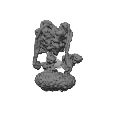
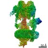
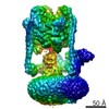
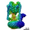
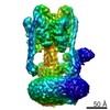
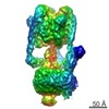
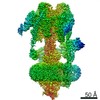
| Entry | Database: EMDB / ID: EMD-0646 | |||||||||
|---|---|---|---|---|---|---|---|---|---|---|
| Title | Saccharomyces cerevisiae V-ATPase Stv1-V1VO State 1 | |||||||||
 Map data Map data | ||||||||||
 Sample Sample |
| |||||||||
 Keywords Keywords | Proton pump / MEMBRANE PROTEIN | |||||||||
| Function / homology |  Function and homology information Function and homology informationvacuole-mitochondrion membrane contact site / cell wall mannoprotein biosynthetic process / ATPase-coupled ion transmembrane transporter activity / proton-transporting V-type ATPase, V1 domain / Insulin receptor recycling / Transferrin endocytosis and recycling / ROS and RNS production in phagocytes / Amino acids regulate mTORC1 / Golgi lumen acidification / proteasome storage granule assembly ...vacuole-mitochondrion membrane contact site / cell wall mannoprotein biosynthetic process / ATPase-coupled ion transmembrane transporter activity / proton-transporting V-type ATPase, V1 domain / Insulin receptor recycling / Transferrin endocytosis and recycling / ROS and RNS production in phagocytes / Amino acids regulate mTORC1 / Golgi lumen acidification / proteasome storage granule assembly / P-type proton-exporting transporter activity / vacuolar transport / vacuolar proton-transporting V-type ATPase, V1 domain / vacuolar proton-transporting V-type ATPase, V0 domain / endosomal lumen acidification / vacuole organization / protein targeting to vacuole / proton-transporting V-type ATPase complex / pexophagy / fungal-type vacuole / vacuolar proton-transporting V-type ATPase complex / vacuolar acidification / fungal-type vacuole membrane / phosphatidylinositol-4-phosphate binding / proton transmembrane transporter activity / proton-transporting ATPase activity, rotational mechanism / intracellular copper ion homeostasis / ATP metabolic process / H+-transporting two-sector ATPase / Neutrophil degranulation / RNA endonuclease activity / proton transmembrane transport / cell periphery / transmembrane transport / endocytosis / intracellular calcium ion homeostasis / cytoplasmic stress granule / late endosome / ATPase binding / protein-containing complex assembly / intracellular iron ion homeostasis / endosome membrane / membrane raft / Golgi membrane / endoplasmic reticulum membrane / Golgi apparatus / ATP hydrolysis activity / ATP binding / membrane / cytoplasm Similarity search - Function | |||||||||
| Biological species |  | |||||||||
| Method | single particle reconstruction / cryo EM / Resolution: 6.6 Å | |||||||||
 Authors Authors | Vasanthakumar T / Bueler SA / Wu D / Beilsten-Edmands V / Robinson CV / Rubinstein JL | |||||||||
| Funding support |  Canada, 1 items Canada, 1 items
| |||||||||
 Citation Citation | Journal: Proc Natl Acad Sci U S A / Year: 2019 Title: Structural comparison of the vacuolar and Golgi V-ATPases from . Authors: Thamiya Vasanthakumar / Stephanie A Bueler / Di Wu / Victoria Beilsten-Edmands / Carol V Robinson / John L Rubinstein /  Abstract: Proton-translocating vacuolar-type ATPases (V-ATPases) are necessary for numerous processes in eukaryotic cells, including receptor-mediated endocytosis, protein maturation, and lysosomal ...Proton-translocating vacuolar-type ATPases (V-ATPases) are necessary for numerous processes in eukaryotic cells, including receptor-mediated endocytosis, protein maturation, and lysosomal acidification. In mammals, V-ATPase subunit isoforms are differentially targeted to various intracellular compartments or tissues, but how these subunit isoforms influence enzyme activity is not clear. In the yeast , isoform diversity is limited to two different versions of the proton-translocating subunit a: Vph1p, which is targeted to the vacuole, and Stv1p, which is targeted to the Golgi apparatus and endosomes. We show that purified V-ATPase complexes containing Vph1p have higher ATPase activity than complexes containing Stv1p and that the relative difference in activity depends on the presence of lipids. We also show that V complexes containing Stv1p could be readily purified without attached V regions. We used this effect to determine structures of the membrane-embedded V region with Stv1p at 3.1-Å resolution, which we compare with a structure of the V region with Vph1p that we determine to 3.2-Å resolution. These maps reveal differences in the surface charge near the cytoplasmic proton half-channel. Both maps also show the presence of bound lipids, as well as regularly spaced densities that may correspond to ergosterol or bound detergent, around the c-ring. | |||||||||
| History |
|
- Structure visualization
| Movie |
Movie viewer |
|---|---|
| Structure viewer | EM map: SurfViewMolmilJmol/JSmol |
| Supplemental images |
- Downloads & links
-EMDB archive
| Map data | emd_0646.map.gz | 31 MB | EMDB map data format | |
|---|---|---|---|---|
| Header (meta data) | emd-0646-v30.xmlemd-0646.xml | 28.8 KB 28.8 KB | Display Display | EMDB header |
| Images |  emd_0646.png emd_0646.png | 29.2 KB | ||
| Filedesc metadata | emd-0646.cif.gz | 8.8 KB | ||
| Archive directory |  http://ftp.pdbj.org/pub/emdb/structures/EMD-0646ftp://ftp.pdbj.org/pub/emdb/structures/EMD-0646 http://ftp.pdbj.org/pub/emdb/structures/EMD-0646ftp://ftp.pdbj.org/pub/emdb/structures/EMD-0646 | HTTPS FTP |
-Related structure data
| Related structure data | 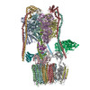 6o7vMC 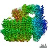 0644C 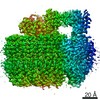 0645C 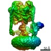 0647C 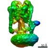 0648C  6o7tC  6o7uC 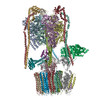 6o7wC 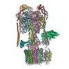 6o7xC C: citing same article ( M: atomic model generated by this map |
|---|---|
| Similar structure data |
-Links
| EMDB pages | EMDB (EBI/PDBe) / EMDataResource |
|---|---|
| Related items in Molecule of the Month |


-Map
| File | Download / File: emd_0646.map.gz / Format: CCP4 / Size: 64 MB / Type: IMAGE STORED AS FLOATING POINT NUMBER (4 BYTES) | ||||||||||||||||||||||||||||||||||||||||||||||||||||||||||||||||||||
|---|---|---|---|---|---|---|---|---|---|---|---|---|---|---|---|---|---|---|---|---|---|---|---|---|---|---|---|---|---|---|---|---|---|---|---|---|---|---|---|---|---|---|---|---|---|---|---|---|---|---|---|---|---|---|---|---|---|---|---|---|---|---|---|---|---|---|---|---|---|
| Projections & slices | Image control
Images are generated by Spider. | ||||||||||||||||||||||||||||||||||||||||||||||||||||||||||||||||||||
| Voxel size | X=Y=Z: 1.45 Å | ||||||||||||||||||||||||||||||||||||||||||||||||||||||||||||||||||||
| Density |
| ||||||||||||||||||||||||||||||||||||||||||||||||||||||||||||||||||||
| Symmetry | Space group: 1 | ||||||||||||||||||||||||||||||||||||||||||||||||||||||||||||||||||||
| Details | EMDB XML:
CCP4 map header:
| ||||||||||||||||||||||||||||||||||||||||||||||||||||||||||||||||||||
 Z (Sec.)
 Y (Row.)
 X (Col.)


-Supplemental data
- Sample components
+Entire : Saccharomyces cerevisiae V-ATPase Stv1-V1VO State 1
+Supramolecule #1: Saccharomyces cerevisiae V-ATPase Stv1-V1VO State 1
+Macromolecule #1: V-type proton ATPase subunit D
+Macromolecule #2: V-type proton ATPase subunit F
+Macromolecule #3: Vacuolar ATP synthase catalytic subunit A
+Macromolecule #4: V-type proton ATPase subunit B
+Macromolecule #5: V-type proton ATPase subunit G
+Macromolecule #6: V-type proton ATPase subunit E
+Macromolecule #7: V-type proton ATPase subunit H
+Macromolecule #8: V-type proton ATPase subunit C
+Macromolecule #9: V-type proton ATPase subunit a, Golgi isoform
+Macromolecule #10: V0 assembly protein 1
+Macromolecule #11: V-type proton ATPase subunit c''
+Macromolecule #12: V-type proton ATPase subunit d
+Macromolecule #13: V-type proton ATPase subunit c
+Macromolecule #14: V-type proton ATPase subunit c'
+Macromolecule #15: V-type proton ATPase subunit e
+Macromolecule #16: Putative protein YPR170W-B
-Experimental details
-Structure determination
| Method | cryo EM |
|---|---|
 Processing Processing | single particle reconstruction |
| Aggregation state | particle |
-Sample preparation
| Buffer | pH: 7.4 |
|---|---|
| Vitrification | Cryogen name: ETHANE-PROPANE |
- Electron microscopy
| Microscope | FEI TECNAI F20 |
|---|---|
| Image recording | Film or detector model: GATAN K2 SUMMIT (4k x 4k) / Detector mode: COUNTING / Average electron dose: 35.0 e/Å2 |
| Electron beam | Acceleration voltage: 200 kV / Electron source:  FIELD EMISSION GUN FIELD EMISSION GUN |
| Electron optics | Illumination mode: FLOOD BEAM / Imaging mode: BRIGHT FIELD |
| Experimental equipment |  Model: Tecnai F20 / Image courtesy: FEI Company |
-Image processing
| Startup model | Type of model: OTHER / Details: Ab initio |
|---|---|
| Final reconstruction | Resolution.type: BY AUTHOR / Resolution: 6.6 Å / Resolution method: FSC 0.143 CUT-OFF / Number images used: 25045 |
| Initial angle assignment | Type: MAXIMUM LIKELIHOOD |
| Final angle assignment | Type: MAXIMUM LIKELIHOOD |
