 Movie
Movie Controller

[English] 日本語
 Yorodumi
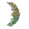
- EMDB-0023: CryoEM structure of the MDA5-dsRNA filament in complex with ADP-AlF4 -
+ Open data

- Basic information
| Entry |  | |||||||||
|---|---|---|---|---|---|---|---|---|---|---|
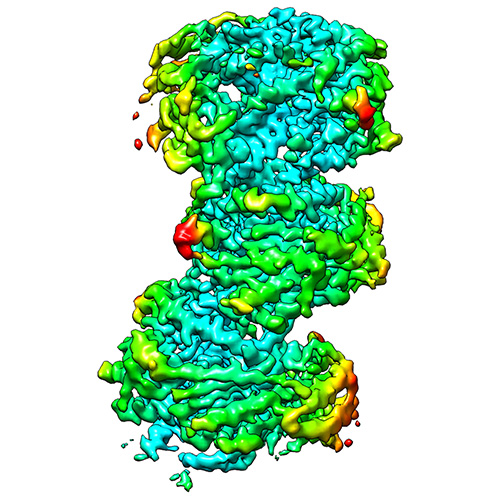
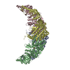
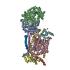
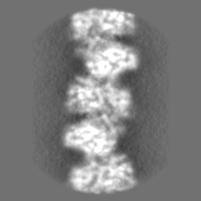
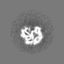
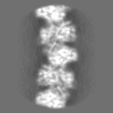
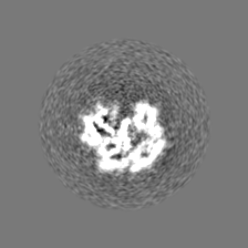
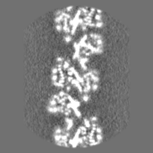
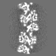
| Title | CryoEM structure of the MDA5-dsRNA filament in complex with ADP-AlF4 | |||||||||
 Map data Map data | Volume used in reconstruction | |||||||||
 Sample Sample |
| |||||||||
 Keywords Keywords | Protein-RNA complex / helical filament / ATPase / innate immune receptor / IMMUNE SYSTEM | |||||||||
| Function / homology |  Function and homology information Function and homology informationMDA-5 signaling pathway / Ub-specific processing proteases / positive regulation of response to cytokine stimulus / type I interferon-mediated signaling pathway / negative regulation of viral genome replication / pattern recognition receptor activity / cellular response to exogenous dsRNA / protein complex oligomerization / positive regulation of interferon-alpha production / protein sumoylation ...MDA-5 signaling pathway / Ub-specific processing proteases / positive regulation of response to cytokine stimulus / type I interferon-mediated signaling pathway / negative regulation of viral genome replication / pattern recognition receptor activity / cellular response to exogenous dsRNA / protein complex oligomerization / positive regulation of interferon-alpha production / protein sumoylation / ribonucleoprotein complex binding / antiviral innate immune response / positive regulation of interferon-beta production / response to virus / cellular response to virus / positive regulation of interleukin-6 production / positive regulation of tumor necrosis factor production / double-stranded RNA binding / defense response to virus / single-stranded RNA binding / RNA helicase activity / RNA helicase / protein domain specific binding / innate immune response / ATP hydrolysis activity / mitochondrion / DNA binding / zinc ion binding / ATP binding / identical protein binding / nucleus / cytoplasm Similarity search - Function | |||||||||
| Biological species |   Pseudomonas savastanoi pv. phaseolicola (bacteria) / Pseudomonas savastanoi pv. phaseolicola (bacteria) /  Pseudomonas phage phi6 (bacteriophage) Pseudomonas phage phi6 (bacteriophage) | |||||||||
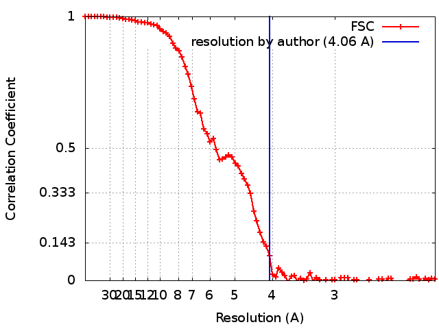
| Method | helical reconstruction / cryo EM / Resolution: 4.06 Å | |||||||||
 Authors Authors | Yu Q / Qu K | |||||||||
| Funding support |  United Kingdom, 2 items United Kingdom, 2 items
| |||||||||
 Citation Citation | Journal: Mol Cell / Year: 2018 Title: Cryo-EM Structures of MDA5-dsRNA Filaments at Different Stages of ATP Hydrolysis. Authors: Qin Yu / Kun Qu / Yorgo Modis / Abstract: Double-stranded RNA (dsRNA) is a potent proinflammatory signature of viral infection. Long cytosolic dsRNA is recognized by MDA5. The cooperative assembly of MDA5 into helical filaments on dsRNA ...Double-stranded RNA (dsRNA) is a potent proinflammatory signature of viral infection. Long cytosolic dsRNA is recognized by MDA5. The cooperative assembly of MDA5 into helical filaments on dsRNA nucleates the assembly of a multiprotein type I interferon signaling platform. Here, we determined cryoelectron microscopy (cryo-EM) structures of MDA5-dsRNA filaments with different helical twists and bound nucleotide analogs at resolutions sufficient to build and refine atomic models. The structures identify the filament-forming interfaces, which encode the dsRNA binding cooperativity and length specificity of MDA5. The predominantly hydrophobic interface contacts confer flexibility, reflected in the variable helical twist within filaments. Mutation of filament-forming residues can result in loss or gain of signaling activity. Each MDA5 molecule spans 14 or 15 RNA base pairs, depending on the twist. Variations in twist also correlate with variations in the occupancy and type of nucleotide in the active site, providing insights on how ATP hydrolysis contributes to MDA5-dsRNA recognition. | |||||||||
| History |
|
- Structure visualization
| Structure viewer | EM map: SurfViewMolmilJmol/JSmol |
|---|---|
| Supplemental images |
- Downloads & links
-EMDB archive
| Map data | emd_0023.map.gz | 5.3 MB | EMDB map data format | |
|---|---|---|---|---|
| Header (meta data) | emd-0023-v30.xmlemd-0023.xml | 24.2 KB 24.2 KB | Display Display | EMDB header |
| FSC (resolution estimation) | emd_0023_fsc.xml | 9.3 KB | Display | FSC data file |
| Images |  emd_0023.png emd_0023.png | 253.2 KB | ||
| Masks | emd_0023_msk_1.map | 42.9 MB | Mask map | |
| Filedesc metadata | emd-0023.cif.gz | 7.6 KB | ||
| Others | emd_0023_half_map_1.map.gzemd_0023_half_map_2.map.gz | 12.8 MB 12.9 MB | ||
| Archive directory |  http://ftp.pdbj.org/pub/emdb/structures/EMD-0023ftp://ftp.pdbj.org/pub/emdb/structures/EMD-0023 http://ftp.pdbj.org/pub/emdb/structures/EMD-0023ftp://ftp.pdbj.org/pub/emdb/structures/EMD-0023 | HTTPS FTP |
-Related structure data
| Related structure data |  6gkhMC  0012C  0024C  0143C 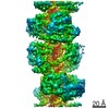 0145C 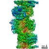 4338C 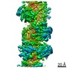 4340C 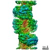 4341C 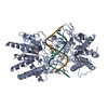 6g19C 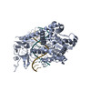 6g1sC 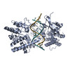 6g1xC 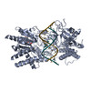 6gjzC 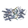 6gkmC 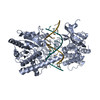 6h61C 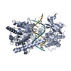 6h66C C: citing same article ( M: atomic model generated by this map |
|---|---|
| Similar structure data | |
| EM raw data | EMPIAR-10211 (Title: mouse MDA5-dsRNA filamemts in complex of 2mM ADP-AlF4 Data size: 195.3 Data #1: mouse MDA5-dsRNA filaments in complex with 2mM ADP-AlF4_data1 [micrographs - single frame] Data #2: mouse MDA5-dsRNA filaments in complex with 2mM ADP-AlF4_data2_rescaled [micrographs - single frame] Data #3: mouse MDA5-dsRNA filaments in complex with 2mM ADP-AlF4_data2 [micrographs - single frame]) |
-Links
| EMDB pages | EMDB (EBI/PDBe) / EMDataResource |
|---|---|
| Related items in Molecule of the Month |


-Map
| File | Download / File: emd_0023.map.gz / Format: CCP4 / Size: 42.9 MB / Type: IMAGE STORED AS FLOATING POINT NUMBER (4 BYTES) | ||||||||||||||||||||||||||||||||||||
|---|---|---|---|---|---|---|---|---|---|---|---|---|---|---|---|---|---|---|---|---|---|---|---|---|---|---|---|---|---|---|---|---|---|---|---|---|---|
| Annotation | Volume used in reconstruction | ||||||||||||||||||||||||||||||||||||
| Projections & slices | Image control
Images are generated by Spider. | ||||||||||||||||||||||||||||||||||||
| Voxel size | X=Y=Z: 1.07 Å | ||||||||||||||||||||||||||||||||||||
| Density |
| ||||||||||||||||||||||||||||||||||||
| Symmetry | Space group: 1 | ||||||||||||||||||||||||||||||||||||
| Details | EMDB XML:
|
 Z (Sec.)
 Y (Row.)
 X (Col.)


-Supplemental data
-Mask #1
| File | emd_0023_msk_1.map | ||||||||||||
|---|---|---|---|---|---|---|---|---|---|---|---|---|---|
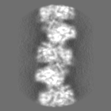
| Projections & Slices |
| ||||||||||||
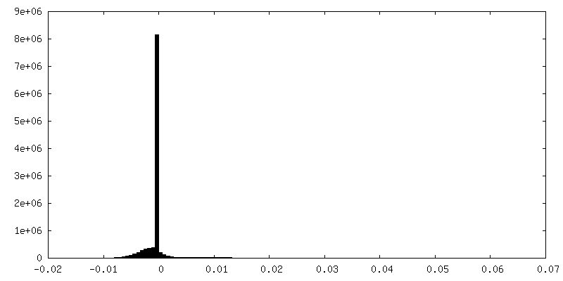
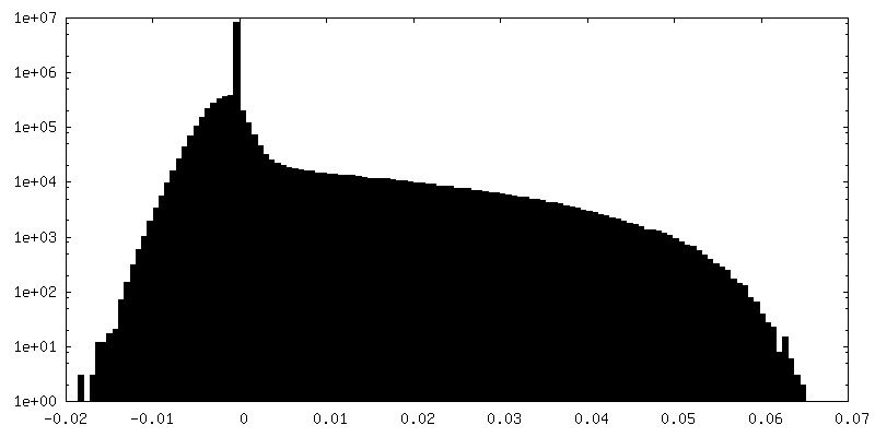
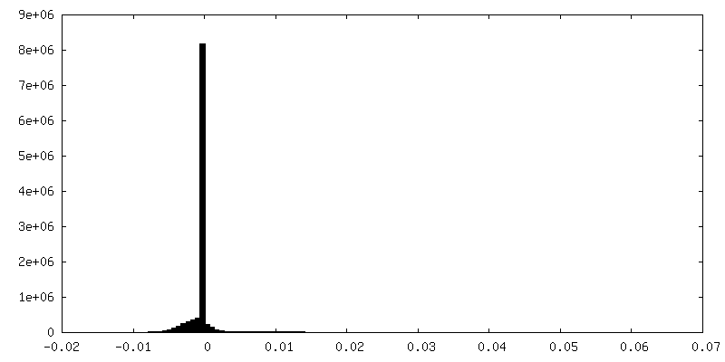
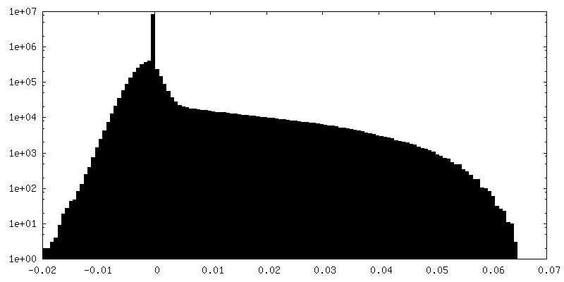
| Density Histograms |


-Half map: Half map 1
| File | emd_0023_half_map_1.map | ||||||||||||
|---|---|---|---|---|---|---|---|---|---|---|---|---|---|
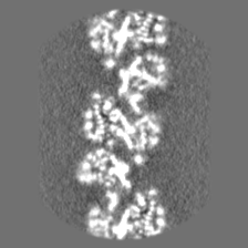
| Annotation | Half map 1 | ||||||||||||
| Projections & Slices |
| ||||||||||||
| Density Histograms |

-Half map: Half map 2
| File | emd_0023_half_map_2.map | ||||||||||||
|---|---|---|---|---|---|---|---|---|---|---|---|---|---|
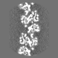
| Annotation | Half map 2 | ||||||||||||
| Projections & Slices |
| ||||||||||||
| Density Histograms |
- Sample components
+Entire : MDA5-dsRNA helical filament in complex with ADP-AlF4
+Supramolecule #1: MDA5-dsRNA helical filament in complex with ADP-AlF4
+Supramolecule #2: MDA5 bound to ADP-AlF4
+Supramolecule #3: Double-stranded RNA from bacteriophage Phi6
+Macromolecule #1: Interferon-induced helicase C domain-containing protein 1
+Macromolecule #2: RNA (5'-R(P*GP*UP*CP*AP*AP*GP*CP*CP*GP*AP*GP*GP*AP*GP*A)-3')
+Macromolecule #3: RNA (5'-R(P*UP*CP*UP*CP*CP*UP*CP*GP*GP*CP*UP*UP*GP*AP*C)-3')
+Macromolecule #4: ADENOSINE-5'-DIPHOSPHATE

+Macromolecule #5: TETRAFLUOROALUMINATE ION

+Macromolecule #6: MAGNESIUM ION
+Macromolecule #7: ZINC ION
-Experimental details
-Structure determination
| Method | cryo EM |
|---|---|
 Processing Processing | helical reconstruction |
| Aggregation state | helical array |
-Sample preparation
| Concentration | 0.5 mg/mL | |||||||||||||||
|---|---|---|---|---|---|---|---|---|---|---|---|---|---|---|---|---|
| Buffer | pH: 7.7 Component:
| |||||||||||||||
| Grid | Model: Quantifoil R1.2/1.3 / Material: GOLD / Mesh: 300 / Support film - Material: CARBON / Support film - topology: HOLEY / Pretreatment - Type: GLOW DISCHARGE / Pretreatment - Time: 60 sec. / Pretreatment - Atmosphere: AIR / Details: 25 mA | |||||||||||||||
| Vitrification | Cryogen name: ETHANE / Chamber humidity: 100 % / Chamber temperature: 277 K / Instrument: FEI VITROBOT MARK IV | |||||||||||||||
| Details | Samples were diluted twofold from 1 mg/ml to 0.5 mg/ml immediately prior to plunge freezing |
- Electron microscopy
| Microscope | FEI TITAN KRIOS |
|---|---|
| Image recording | Film or detector model: FEI FALCON III (4k x 4k) / Detector mode: COUNTING / Digitization - Dimensions - Width: 4096 pixel / Digitization - Dimensions - Height: 4096 pixel / Average electron dose: 29.85 e/Å2 |
| Electron beam | Acceleration voltage: 300 kV / Electron source:  FIELD EMISSION GUN FIELD EMISSION GUN |
| Electron optics | Calibrated magnification: 75000 / Illumination mode: FLOOD BEAM / Imaging mode: BRIGHT FIELD / Nominal defocus max: -2.7 µm / Nominal defocus min: -1.8 µm |
| Sample stage | Specimen holder model: FEI TITAN KRIOS AUTOGRID HOLDER / Cooling holder cryogen: NITROGEN |
| Experimental equipment |  Model: Titan Krios / Image courtesy: FEI Company |
+Image processing
-Atomic model buiding 1
| Refinement | Space: REAL / Protocol: FLEXIBLE FIT / Overall B value: 175 / Target criteria: Cross-correlation coefficient |
|---|---|
| Output model | PDB-6gkh: |
