Niko W Vlahakis / Cameron W Flowers / Mengting Liu / Matthew Agdanowski / Samuel Johnson / Jacob A Summers / Catherine Keyser / Phoebe Russell / Samuel Rose / Julien Orlans / Nima Adhami / Yu Chen / Michael R Sawaya / Shibom Basu / Daniele de Sanctis / Soichi Wakatsuki / Hosea M Nelson / Joseph A Loo / Yi Tang / Jose A Rodriguez /
PubMed Abstract
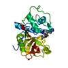
With the goal of accelerating the discovery of small molecule-protein complexes, we leverage fast, low-dose, event based electron counting microcrystal electron diffraction (MicroED) data collection ...With the goal of accelerating the discovery of small molecule-protein complexes, we leverage fast, low-dose, event based electron counting microcrystal electron diffraction (MicroED) data collection and native mass spectrometry. This approach resolves structures of the epoxide-based cysteine protease inhibitor, and natural product, E-64, and its biosynthetic analogs bound to the model cysteine protease, papain. The combined structural power of MicroED and the analytical capabilities of native mass spectrometry (ED-MS) allows assignment of papain structures bound to E-64-like ligands with data obtained from crystal slurries soaked with mixtures of known inhibitors, and crude biosynthetic reactions. ED-MS further discriminates the highest-affinity ligand soaked into microcrystals from a broad inhibitor cocktail, and identifies multiple similarly high-affinity ligands soaked into microcrystals simultaneously. This extends to libraries of printed ligands dispensed directly onto TEM grids and later soaked with papain microcrystal slurries. ED-MS identifies papain binding to its preferred natural products, by showing that two analogues of E-64 outcompete others in binding to papain crystals, and by detecting papain bound to E-64 and an analogue from crude biosynthetic reactions, without purification. This illustrates the utility of ED-MS for natural product ligand discovery and for structure-based screening of small molecule binders to macromolecular targets.
PDB-9n9d: MicroED structure of papain co-crystallized with E-64C Method: ELECTRON CRYSTALLOGRAPHY / Resolution: 2.2 Å
PDB-9nae: MicroED structure of papain co-crystallized with E-64 Method: ELECTRON CRYSTALLOGRAPHY / Resolution: 2.3 Å
PDB-9nag: MicroED structure of the apo-form of papain Method: ELECTRON CRYSTALLOGRAPHY / Resolution: 2.5 Å
PDB-9nao: MicroED structure of papain complexed with natural product E64-A65 Method: ELECTRON CRYSTALLOGRAPHY / Resolution: 2.5 Å
PDB-9nar: MicroED structure of papain microcrystals soaked with E-64 for 10 minutes Method: ELECTRON CRYSTALLOGRAPHY / Resolution: 2.5 Å
PDB-9nat: X-ray diffraction structure of papain co-crystallized with leupeptin Method: X-RAY DIFFRACTION / Resolution: 1.6 Å
PDB-9nax: MicroED structure of the papain-E-64 complex from microcrystals soaked with crude biosynthetic reaction mixture Method: ELECTRON CRYSTALLOGRAPHY / Resolution: 2.3 Å
PDB-9nay: MicroED structure of papain complexed with natural product E-64-A65 from microcrystals soaked in crude biosynthetic reaction mixture Method: ELECTRON CRYSTALLOGRAPHY / Resolution: 2.5 Å
PDB-9nb2: X-ray diffraction structure of papain soaked with E-64 Method: X-RAY DIFFRACTION / Resolution: 1.5 Å
PDB-9nb4: Serial synchrotron X-ray diffraction structure of papain microcrystals soaked with natural product E-64-A65 Method: X-RAY DIFFRACTION / Resolution: 1.8 Å
PDB-9nb7: Serial synchrotron X-ray diffraction structure of papain microcrystals soaked with natural product E405 Method: X-RAY DIFFRACTION / Resolution: 1.8 Å
PDB-9nbf: Serial synchrotron X-ray diffraction structure of papain microcrystals soaked with E-64 Method: X-RAY DIFFRACTION / Resolution: 1.8 Å
PDB-9nbj: Serial synchrotron X-ray diffraction structure of papain microcrystals soaked with E-64C Method: X-RAY DIFFRACTION / Resolution: 1.8 Å
PDB-9nbk: Serial synchrotron X-ray diffraction structure of papain microcrystals soaked with E-64D Method: X-RAY DIFFRACTION / Resolution: 1.8 Å
PDB-9nbn: Serial synchrotron X-ray diffraction structure of papain microcrystals soaked with a mixture of E-64, E-64C, and E-64D Method: X-RAY DIFFRACTION / Resolution: 1.8 Å
PDB-9nbp: MicroED structure of the papain-E-64 complex from microcrystals mixed on-grid with microarrayed ligand Method: ELECTRON CRYSTALLOGRAPHY / Resolution: 2.8 Å
PDB-9nbq: MicroED structure of papain co-crystallized with E-64D Method: ELECTRON CRYSTALLOGRAPHY / Resolution: 2.3 Å
PDB-9nc1: MicroED structure of papain-E-64 complex from microcrystals soaked with protease inhibitor cocktail Method: ELECTRON CRYSTALLOGRAPHY / Resolution: 2.4 Å
PDB-9nca: MicroED structure of microcrystals soaked with a mixture of E-64, E-64C, and E-64D Method: ELECTRON CRYSTALLOGRAPHY / Resolution: 2.5 Å
In the structure databanks used in Yorodumi, some data are registered as the other names, "COVID-19 virus" and "2019-nCoV". Here are the details of the virus and the list of structure data.
Jan 31, 2019. EMDB accession codes are about to change! (news from PDBe EMDB page)
EMDB accession codes are about to change! (news from PDBe EMDB page)
The allocation of 4 digits for EMDB accession codes will soon come to an end. Whilst these codes will remain in use, new EMDB accession codes will include an additional digit and will expand incrementally as the available range of codes is exhausted. The current 4-digit format prefixed with “EMD-” (i.e. EMD-XXXX) will advance to a 5-digit format (i.e. EMD-XXXXX), and so on. It is currently estimated that the 4-digit codes will be depleted around Spring 2019, at which point the 5-digit format will come into force.
The EM Navigator/Yorodumi systems omit the EMD- prefix.
Related info.:Q: What is EMD? / ID/Accession-code notation in Yorodumi/EM Navigator
 Movie
Movie Controller
 Structure viewers
 About Yorodumi Papers


 Authors

 External links


 Keywords
 carica papaya (papaya)