 Movie
Movie Controller

[English] 日本語
 Yorodumi
- PDB-9nax: MicroED structure of the papain-E-64 complex from microcrystals s... -
+ Open data

- Basic information
| Entry | Database: PDB / ID: 9nax | ||||||||||||
|---|---|---|---|---|---|---|---|---|---|---|---|---|---|
| Title | MicroED structure of the papain-E-64 complex from microcrystals soaked with crude biosynthetic reaction mixture | ||||||||||||
 Components Components | Papain | ||||||||||||
 Keywords Keywords | HYDROLASE / Inhibitor / Complex / Protease / MicroED | ||||||||||||
| Function / homology |  Function and homology information Function and homology informationpapain / serpin family protein binding / cysteine-type peptidase activity / proteolysis Similarity search - Function | ||||||||||||
| Biological species |  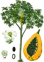 Carica papaya (papaya) Carica papaya (papaya) | ||||||||||||
| Method | ELECTRON CRYSTALLOGRAPHY / electron crystallography /  MOLECULAR REPLACEMENT / cryo EM / Resolution: 2.3 Å MOLECULAR REPLACEMENT / cryo EM / Resolution: 2.3 Å | ||||||||||||
 Authors Authors | Vlahakis, N.W. / Rodriguez, J.A. | ||||||||||||
| Funding support |  United States, 3items United States, 3items
| ||||||||||||
 Citation Citation | Journal: bioRxiv / Year: 2025 Title: Combining MicroED and native mass spectrometry for structural discovery of enzyme-biosynthetic inhibitor complexes. Authors: Niko W Vlahakis / Cameron W Flowers / Mengting Liu / Matthew Agdanowski / Samuel Johnson / Jacob A Summers / Catherine Keyser / Phoebe Russell / Samuel Rose / Julien Orlans / Nima Adhami / ...Authors: Niko W Vlahakis / Cameron W Flowers / Mengting Liu / Matthew Agdanowski / Samuel Johnson / Jacob A Summers / Catherine Keyser / Phoebe Russell / Samuel Rose / Julien Orlans / Nima Adhami / Yu Chen / Michael R Sawaya / Shibom Basu / Daniele de Sanctis / Soichi Wakatsuki / Hosea M Nelson / Joseph A Loo / Yi Tang / Jose A Rodriguez /  Abstract: With the goal of accelerating the discovery of small molecule-protein complexes, we leverage fast, low-dose, event based electron counting microcrystal electron diffraction (MicroED) data collection ...With the goal of accelerating the discovery of small molecule-protein complexes, we leverage fast, low-dose, event based electron counting microcrystal electron diffraction (MicroED) data collection and native mass spectrometry. This approach resolves structures of the epoxide-based cysteine protease inhibitor, and natural product, E-64, and its biosynthetic analogs bound to the model cysteine protease, papain. The combined structural power of MicroED and the analytical capabilities of native mass spectrometry (ED-MS) allows assignment of papain structures bound to E-64-like ligands with data obtained from crystal slurries soaked with mixtures of known inhibitors, and crude biosynthetic reactions. ED-MS further discriminates the highest-affinity ligand soaked into microcrystals from a broad inhibitor cocktail, and identifies multiple similarly high-affinity ligands soaked into microcrystals simultaneously. This extends to libraries of printed ligands dispensed directly onto TEM grids and later soaked with papain microcrystal slurries. ED-MS identifies papain binding to its preferred natural products, by showing that two analogues of E-64 outcompete others in binding to papain crystals, and by detecting papain bound to E-64 and an analogue from crude biosynthetic reactions, without purification. This illustrates the utility of ED-MS for natural product ligand discovery and for structure-based screening of small molecule binders to macromolecular targets. | ||||||||||||
| History |
|
- Structure visualization
| Structure viewer | Molecule: MolmilJmol/JSmol |
|---|
- Downloads & links
-Download
| PDBx/mmCIF format | 9nax.cif.gz | 56.2 KB | Display | PDBx/mmCIF format |
|---|---|---|---|---|
| PDB format | pdb9nax.ent.gz | 38.8 KB | Display | PDB format |
| PDBx/mmJSON format | 9nax.json.gz | Tree view | PDBx/mmJSON format | |
| Others |  Other downloads Other downloads |
-Validation report
| Arichive directory | https://data.pdbj.org/pub/pdb/validation_reports/na/9naxftp://data.pdbj.org/pub/pdb/validation_reports/na/9nax | HTTPS FTP |
|---|
-Related structure data
| Related structure data |  9n9dC  9naeC  9nagC  9naoC  9narC  9natC  9nayC  9nb2C  9nb4C  9nb7C 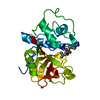 9nbfC  9nbjC  9nbkC  9nbnC C: citing same article ( |
|---|---|
| Similar structure data |
-Links
 PDBj
PDBj
- Assembly
| Deposited unit | 
| ||||||||
|---|---|---|---|---|---|---|---|---|---|
| 1 |
| ||||||||
| Unit cell |
|
-Components
| #1: Protein | Mass: 23452.301 Da / Num. of mol.: 1 / Source method: isolated from a natural source / Source: (natural) Carica papaya (papaya) / References: UniProt: P00784, papain |
|---|---|
| #2: Chemical | ChemComp-E64 /   Mass: 360.429 Da / Num. of mol.: 1 / Source method: obtained synthetically / Formula: C15H30N5O5 / Feature type: SUBJECT OF INVESTIGATION Mass: 360.429 Da / Num. of mol.: 1 / Source method: obtained synthetically / Formula: C15H30N5O5 / Feature type: SUBJECT OF INVESTIGATION |
| #3: Water | ChemComp-HOH /  Mass: 18.015 Da / Num. of mol.: 17 / Source method: isolated from a natural source / Formula: H2O Mass: 18.015 Da / Num. of mol.: 17 / Source method: isolated from a natural source / Formula: H2O |
| Has ligand of interest | Y |
| Has protein modification | Y |
-Experimental details
-Experiment
| Experiment | Method: ELECTRON CRYSTALLOGRAPHY |
|---|---|
| EM experiment | Aggregation state: 3D ARRAY / 3D reconstruction method: electron crystallography |
- Sample preparation
| Component | Name: Papain-E64-C complex / Type: CELL / Entity ID: #1 / Source: NATURAL |
|---|---|
| Source (natural) | Organism: Carica papaya (papaya) |
| Buffer solution | pH: 7 |
| Specimen | Embedding applied: NO / Shadowing applied: NO / Staining applied: NO / Vitrification applied: YES |
| Vitrification | Cryogen name: ETHANE |
-Data collection
| Experimental equipment |  Model: Tecnai F20 / Image courtesy: FEI Company |
|---|---|
| Microscopy | Model: TFS TALOS F200C |
| Electron gun | Electron source: FIELD EMISSION GUN / Accelerating voltage: 200 kV / Illumination mode: FLOOD BEAM |
| Electron lens | Mode: DIFFRACTION / Nominal defocus max: 10000 nm / Nominal defocus min: 10000 nm / C2 aperture diameter: 70 µm |
| Specimen holder | Cryogen: NITROGEN Specimen holder model: GATAN 626 SINGLE TILT LIQUID NITROGEN CRYO TRANSFER HOLDER Temperature (max): 100 K / Temperature (min): 100 K |
| Image recording | Electron dose: 0.045 e/Å2 / Film or detector model: DIRECT ELECTRON APOLLO (4k x 4k) |
| EM diffraction shell | Resolution: 2.3→2.4 Å / Fourier space coverage: 87.6 % / Multiplicity: 8.3 / Num. of structure factors: 958 / Phase residual: 30.3 ° |
| EM diffraction stats | Fourier space coverage: 87 % / High resolution: 2.3 Å / Num. of intensities measured: 69072 / Num. of structure factors: 8337 / Phase error rejection criteria: NULL / Rmerge: 28 |
- Processing
| Software |
| |||||||||||||||||||||||||||||||||||||||||||||||||
|---|---|---|---|---|---|---|---|---|---|---|---|---|---|---|---|---|---|---|---|---|---|---|---|---|---|---|---|---|---|---|---|---|---|---|---|---|---|---|---|---|---|---|---|---|---|---|---|---|---|---|
| EM 3D crystal entity | ∠α: 90 ° / ∠β: 90 ° / ∠γ: 90 ° / A: 41.85 Å / B: 48.63 Å / C: 99.87 Å / Space group name: P212121 / Space group num: 19 | |||||||||||||||||||||||||||||||||||||||||||||||||
| CTF correction | Type: NONE | |||||||||||||||||||||||||||||||||||||||||||||||||
| 3D reconstruction | Resolution: 2.3 Å / Resolution method: DIFFRACTION PATTERN/LAYERLINES / Symmetry type: 3D CRYSTAL | |||||||||||||||||||||||||||||||||||||||||||||||||
| Atomic model building | PDB-ID: 9PAP Accession code: 9PAP / Source name: PDB / Type: experimental model | |||||||||||||||||||||||||||||||||||||||||||||||||
| Refinement | Method to determine structure: MOLECULAR REPLACEMENT / Resolution: 2.3→49.93 Å / SU ML: 0.29 / Cross valid method: FREE R-VALUE / σ(F): 1.35 / Phase error: 24.17 / Stereochemistry target values: ML
| |||||||||||||||||||||||||||||||||||||||||||||||||
| Solvent computation | Shrinkage radii: 0.9 Å / VDW probe radii: 1.1 Å / Solvent model: FLAT BULK SOLVENT MODEL | |||||||||||||||||||||||||||||||||||||||||||||||||
| Refine LS restraints |
| |||||||||||||||||||||||||||||||||||||||||||||||||
| LS refinement shell |
|

