 Movie
Movie Controller
 Structure viewers
 About Yorodumi Papers


+Search query
-Structure paper
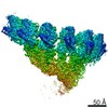
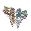
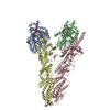
| Title | CM1-driven assembly and activation of yeast γ-tubulin small complex underlies microtubule nucleation. |
|---|---|
| Journal, issue, pages | Elife, Vol. 10, Year 2021 |
| Publish date | May 5, 2021 |
 Authors Authors | Axel F Brilot / Andrew S Lyon / Alex Zelter / Shruthi Viswanath / Alison Maxwell / Michael J MacCoss / Eric G Muller / Andrej Sali / Trisha N Davis / David A Agard /  |
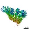
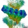
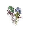
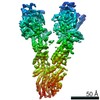
| PubMed Abstract | Microtubule (MT) nucleation is regulated by the γ-tubulin ring complex (γTuRC), conserved from yeast to humans. In , γTuRC is composed of seven identical γ-tubulin small complex (γTuSC) sub- ...Microtubule (MT) nucleation is regulated by the γ-tubulin ring complex (γTuRC), conserved from yeast to humans. In , γTuRC is composed of seven identical γ-tubulin small complex (γTuSC) sub-assemblies, which associate helically to template MT growth. γTuRC assembly provides a key point of regulation for the MT cytoskeleton. Here, we combine crosslinking mass spectrometry, X-ray crystallography, and cryo-EM structures of both monomeric and dimeric γTuSCs, and open and closed helical γTuRC assemblies in complex with Spc110p to elucidate the mechanisms of γTuRC assembly. γTuRC assembly is substantially aided by the evolutionarily conserved CM1 motif in Spc110p spanning a pair of adjacent γTuSCs. By providing the highest resolution and most complete views of any γTuSC assembly, our structures allow phosphorylation sites to be mapped, surprisingly suggesting that they are mostly inhibitory. A comparison of our structures with the CM1 binding site in the human γTuRC structure at the interface between GCP2 and GCP6 allows for the interpretation of significant structural changes arising from CM1 helix binding to metazoan γTuRC. |
 External links External links | Elife / PubMed:33949948 / PubMed Central |
| Methods | EM (helical sym.) / EM (single particle) / X-ray diffraction |
| Resolution | 2.00001856449 - 4.45 Å |
| Structure data | EMDB-23635, PDB-7m2w: EMDB-23636, PDB-7m2x: EMDB-23637, PDB-7m2y: EMDB-23638, PDB-7m2z: 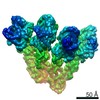 EMDB-23639: 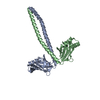 PDB-7m3p: |
| Chemicals |  ChemComp-GTP:  ChemComp-GDP:  ChemComp-HOH: |
| Source |
|
 Keywords Keywords | CELL CYCLE / microtubule nucleation |


 homo sapiens (human)