Movie
Movie Controller
 Structure viewers
 About Yorodumi Papers


+Search query
-Structure paper
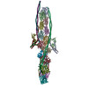
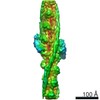
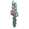
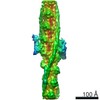
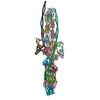
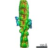
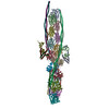
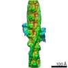
| Title | The structure of the native cardiac thin filament at systolic Ca levels. |
|---|---|
| Journal, issue, pages | Proc Natl Acad Sci U S A, Vol. 118, Issue 13, Year 2021 |
| Publish date | Mar 30, 2021 |
 Authors Authors | Cristina M Risi / Ian Pepper / Betty Belknap / Maicon Landim-Vieira / Howard D White / Kelly Dryden / Jose R Pinto / P Bryant Chase / Vitold E Galkin /  |
| PubMed Abstract | Every heartbeat relies on cyclical interactions between myosin thick and actin thin filaments orchestrated by rising and falling Ca levels. Thin filaments are comprised of two actin strands, each ...Every heartbeat relies on cyclical interactions between myosin thick and actin thin filaments orchestrated by rising and falling Ca levels. Thin filaments are comprised of two actin strands, each harboring equally separated troponin complexes, which bind Ca to move tropomyosin cables away from the myosin binding sites and, thus, activate systolic contraction. Recently, structures of thin filaments obtained at low (pCa ∼9) or high (pCa ∼3) Ca levels revealed the transition between the Ca-free and Ca-bound states. However, in working cardiac muscle, Ca levels fluctuate at intermediate values between pCa ∼6 and pCa ∼7. The structure of the thin filament at physiological Ca levels is unknown. We used cryoelectron microscopy and statistical analysis to reveal the structure of the cardiac thin filament at systolic pCa = 5.8. We show that the two strands of the thin filament consist of a mixture of regulatory units, which are composed of Ca-free, Ca-bound, or mixed (e.g., Ca free on one side and Ca bound on the other side) troponin complexes. We traced troponin complex conformations along and across individual thin filaments to directly determine the structural composition of the cardiac native thin filament at systolic Ca levels. We demonstrate that the two thin filament strands are activated stochastically with short-range cooperativity evident only on one of the two strands. Our findings suggest a mechanism by which cardiac muscle is regulated by narrow range Ca fluctuations. |
 External links External links | Proc Natl Acad Sci U S A / PubMed:33753506 / PubMed Central |
| Methods | EM (single particle) |
| Resolution | 7.8 - 8.3 Å |
| Structure data | EMDB-22964, PDB-7ko4: EMDB-22965, PDB-7ko5: EMDB-22966, PDB-7ko7: EMDB-22978, PDB-7kon: EMDB-22981, PDB-7kor: |
| Source |
|
 Keywords Keywords | CONTRACTILE PROTEIN / tropomyosin / troponin / actin / TF / cardiac |