 Movie
Movie Controller

[English] 日本語
 Yorodumi
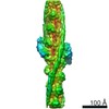
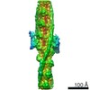
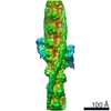
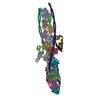
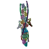
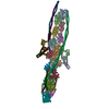
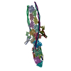
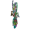
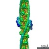
- PDB-7kon: Structure of upper Tn Ca2+ free (rotated) and lower Tn Ca2+ bound... -
+ Open data

- Basic information
| Entry | Database: PDB / ID: 7kon | ||||||||||||
|---|---|---|---|---|---|---|---|---|---|---|---|---|---|
| Title | Structure of upper Tn Ca2+ free (rotated) and lower Tn Ca2+ bound cardiac native thin filament at pCa=5.8 | ||||||||||||
 Components Components |
| ||||||||||||
 Keywords Keywords | CONTRACTILE PROTEIN / actin / tropomyosin / troponin / TF | ||||||||||||
| Biological species |  | ||||||||||||
| Method | ELECTRON MICROSCOPY / single particle reconstruction / cryo EM / Resolution: 8.1 Å | ||||||||||||
 Authors Authors | Galkin, V.E. / Risi, C.M. | ||||||||||||
| Funding support |  United States, 3items United States, 3items
| ||||||||||||
 Citation Citation | Journal: Proc Natl Acad Sci U S A / Year: 2021 Title: The structure of the native cardiac thin filament at systolic Ca levels. Authors: Cristina M Risi / Ian Pepper / Betty Belknap / Maicon Landim-Vieira / Howard D White / Kelly Dryden / Jose R Pinto / P Bryant Chase / Vitold E Galkin / Abstract: Every heartbeat relies on cyclical interactions between myosin thick and actin thin filaments orchestrated by rising and falling Ca levels. Thin filaments are comprised of two actin strands, each ...Every heartbeat relies on cyclical interactions between myosin thick and actin thin filaments orchestrated by rising and falling Ca levels. Thin filaments are comprised of two actin strands, each harboring equally separated troponin complexes, which bind Ca to move tropomyosin cables away from the myosin binding sites and, thus, activate systolic contraction. Recently, structures of thin filaments obtained at low (pCa ∼9) or high (pCa ∼3) Ca levels revealed the transition between the Ca-free and Ca-bound states. However, in working cardiac muscle, Ca levels fluctuate at intermediate values between pCa ∼6 and pCa ∼7. The structure of the thin filament at physiological Ca levels is unknown. We used cryoelectron microscopy and statistical analysis to reveal the structure of the cardiac thin filament at systolic pCa = 5.8. We show that the two strands of the thin filament consist of a mixture of regulatory units, which are composed of Ca-free, Ca-bound, or mixed (e.g., Ca free on one side and Ca bound on the other side) troponin complexes. We traced troponin complex conformations along and across individual thin filaments to directly determine the structural composition of the cardiac native thin filament at systolic Ca levels. We demonstrate that the two thin filament strands are activated stochastically with short-range cooperativity evident only on one of the two strands. Our findings suggest a mechanism by which cardiac muscle is regulated by narrow range Ca fluctuations. | ||||||||||||
| History |
|
- Structure visualization
| Movie |
 Movie viewer Movie viewer |
|---|---|
| Structure viewer | Molecule: MolmilJmol/JSmol |
- Downloads & links
-Download
| PDBx/mmCIF format | 7kon.cif.gz | 1.2 MB | Display | PDBx/mmCIF format |
|---|---|---|---|---|
| PDB format | pdb7kon.ent.gz | 1 MB | Display | PDB format |
| PDBx/mmJSON format | 7kon.json.gz | Tree view | PDBx/mmJSON format | |
| Others |  Other downloads Other downloads |
-Validation report
| Arichive directory | https://data.pdbj.org/pub/pdb/validation_reports/ko/7konftp://data.pdbj.org/pub/pdb/validation_reports/ko/7kon | HTTPS FTP |
|---|
-Related structure data
| Related structure data | 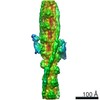 22978MC 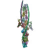 7ko4C 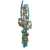 7ko5C 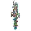 7ko7C  7korC M: map data used to model this data C: citing same article ( |
|---|---|
| Similar structure data |


-Links
 PDBj
PDBj
- Assembly
| Deposited unit | 
|
|---|---|
| 1 |
|
-Components
| #1: Protein | Mass: 41862.613 Da / Num. of mol.: 15 / Source method: isolated from a natural source / Source: (natural) #2: Protein | Mass: 32921.773 Da / Num. of mol.: 8 / Source method: isolated from a natural source / Source: (natural) #3: Protein | Mass: 23023.039 Da / Num. of mol.: 2 / Source method: isolated from a natural source / Source: (natural) #4: Protein | Mass: 19639.691 Da / Num. of mol.: 2 / Source method: isolated from a natural source / Source: (natural) #5: Protein | Mass: 18288.287 Da / Num. of mol.: 2 / Source method: isolated from a natural source / Source: (natural) Sequence details | The resolution within the thin filament is not enough to rebuild the model using the porcine ...The resolution within the thin filament is not enough to rebuild the model using the porcine sequence, so previously determined structures of equivalent proteins from different organisms were fit to the map | |
|---|
-Experimental details
-Experiment
| Experiment | Method: ELECTRON MICROSCOPY |
|---|---|
| EM experiment | Aggregation state: FILAMENT / 3D reconstruction method: single particle reconstruction |
- Sample preparation
| Component | Name: Structure of Ca2+ free / Ca2+ bound cardiac native thin filament at pCa=5.8 mode 2 Type: COMPLEX / Entity ID: all / Source: NATURAL |
|---|---|
| Molecular weight | Experimental value: NO |
| Source (natural) | Organism: |
| Buffer solution | pH: 7 |
| Specimen | Embedding applied: NO / Shadowing applied: NO / Staining applied: NO / Vitrification applied: YES / Details: non-helical filament |
| Specimen support | Grid material: COPPER / Grid mesh size: 300 divisions/in. / Grid type: PELCO Ultrathin Carbon with Lacey Carbon |
| Vitrification | Instrument: FEI VITROBOT MARK IV / Cryogen name: ETHANE / Humidity: 100 % / Chamber temperature: 283 K |
- Electron microscopy imaging
| Microscopy | Model: FEI TITAN |
|---|---|
| Electron gun | Electron source:  FIELD EMISSION GUN / Accelerating voltage: 300 kV / Illumination mode: FLOOD BEAM FIELD EMISSION GUN / Accelerating voltage: 300 kV / Illumination mode: FLOOD BEAM |
| Electron lens | Mode: BRIGHT FIELD |
| Specimen holder | Cryogen: NITROGEN / Specimen holder model: FEI TITAN KRIOS AUTOGRID HOLDER |
| Image recording | Electron dose: 34 e/Å2 / Film or detector model: GATAN K3 (6k x 4k) Details: Images were collected in movie-mode - 40 subframes at 0.85e/A^2 |
- Processing
| Software | Name: PHENIX / Version: 1.15.2_3472: / Classification: refinement | ||||||||||||||||||||||||||||||||
|---|---|---|---|---|---|---|---|---|---|---|---|---|---|---|---|---|---|---|---|---|---|---|---|---|---|---|---|---|---|---|---|---|---|
| EM software |
| ||||||||||||||||||||||||||||||||
| Image processing | Details: images with energy filter enabled at super-resolution mode | ||||||||||||||||||||||||||||||||
| CTF correction | Type: PHASE FLIPPING AND AMPLITUDE CORRECTION | ||||||||||||||||||||||||||||||||
| 3D reconstruction | Resolution: 8.1 Å / Resolution method: FSC 0.143 CUT-OFF / Num. of particles: 10593 / Algorithm: BACK PROJECTION / Details: The map was filtered to 11A / Symmetry type: POINT | ||||||||||||||||||||||||||||||||
| Atomic model building | Protocol: FLEXIBLE FIT / Space: REAL | ||||||||||||||||||||||||||||||||
| Atomic model building |
|


