 Movie
Movie Controller
 Structure viewers
 About Yorodumi Papers


+Search query
-Structure paper
| Title | Cryo-EM structures reveal two distinct conformational states in a picornavirus cell entry intermediate. |
|---|---|
| Journal, issue, pages | PLoS Pathog, Vol. 16, Issue 9, Page e1008920, Year 2020 |
| Publish date | Sep 30, 2020 |
 Authors Authors | Pranav N M Shah / David J Filman / Krishanthi S Karunatilaka / Emma L Hesketh / Elisabetta Groppelli / Mike Strauss / James M Hogle /    |
| PubMed Abstract | The virions of enteroviruses such as poliovirus undergo a global conformational change after binding to the cellular receptor, characterized by a 4% expansion, and by the opening of holes at the two ...The virions of enteroviruses such as poliovirus undergo a global conformational change after binding to the cellular receptor, characterized by a 4% expansion, and by the opening of holes at the two and quasi-three-fold symmetry axes of the capsid. The resultant particle is called a 135S particle or A-particle and is thought to be on the pathway to a productive infection. Previously published studies have concluded that the membrane-interactive peptides, namely VP4 and the N-terminus of VP1, are irreversibly externalized in the 135S particle. However, using established protocols to produce the 135S particle, and single particle cryo-electron microscopy methods, we have identified at least two unique states that we call the early and late 135S particle. Surprisingly, only in the "late" 135S particles have detectable levels of the VP1 N-terminus been trapped outside the capsid. Moreover, we observe a distinct density inside the capsid that can be accounted for by VP4 that remains associated with the genome. Taken together our results conclusively demonstrate that the 135S particle is not a unique conformation, but rather a family of conformations that could exist simultaneously. |
 External links External links | PLoS Pathog / PubMed:32997730 / PubMed Central |
| Methods | EM (single particle) |
| Resolution | 2.9 - 3.6 Å |
| Structure data | EMDB-20275, PDB-6p9o: EMDB-20276, PDB-6p9w: EMDB-20469, PDB-6psz: 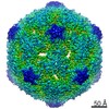 EMDB-20474: EMDB-20546, PDB-6q0b: |
| Source |
|
 Keywords Keywords | VIRUS / Virus-antibody complex / poliovirus / cell-entry intermediate / expanded virus / a-particle / VIRUS/IMMUNE SYSTEM / VIRUS-IMMUNE SYSTEM complex |
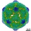
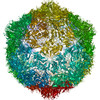
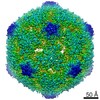

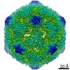
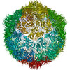
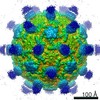
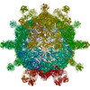

 poliovirus type 1 (strain mahoney)
