- Database of articles cited by EMDB/PDB/SASBDB data -
+
Search query
-
Structure paper
Title
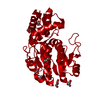
Structure and function of L-threonine-3-dehydrogenase from the parasitic protozoan Trypanosoma brucei revealed by X-ray crystallography and geometric simulations.
PDB-5k4q: Three-dimensional structure of L-threonine 3-dehydrogenase from Trypanosoma brucei bound to NAD+ refined to 2.3 angstroms Method: X-RAY DIFFRACTION / Resolution: 2.3 Å
PDB-5k4t: Three-dimensional structure of L-threonine 3-dehydrogenase from Trypanosoma brucei refined to 2.1 angstroms Method: X-RAY DIFFRACTION / Resolution: 2.1 Å
PDB-5k4u: Three-dimensional structure of L-threonine 3-dehydrogenase from Trypanosoma brucei showing different active site loop conformations between dimer subunits, refined to 1.9 angstroms Method: X-RAY DIFFRACTION / Resolution: 1.9 Å
PDB-5k4v: Three-dimensional structure of L-threonine 3-dehydrogenase from Trypanosoma brucei bound to NAD+ refined to 2.2 angstroms Method: X-RAY DIFFRACTION / Resolution: 2.2 Å
PDB-5k4w: Three-dimensional structure of L-threonine 3-dehydrogenase from Trypanosoma brucei bound to NADH and L-threonine refined to 1.72 angstroms Method: X-RAY DIFFRACTION / Resolution: 1.72 Å
PDB-5k4y: Three-dimensional structure of L-threonine 3-dehydrogenase from Trypanosoma brucei refined to 1.77 angstroms Method: X-RAY DIFFRACTION / Resolution: 1.77 Å
PDB-5k50: Three-dimensional structure of L-threonine 3-dehydrogenase from Trypanosoma brucei bound to NAD+ and L-allo-threonine refined to 2.23 angstroms Method: X-RAY DIFFRACTION / Resolution: 2.26 Å
PDB-5l9a: L-threonine dehydrogenase from trypanosoma brucei. Method: X-RAY DIFFRACTION / Resolution: 1.45 Å
PDB-5lc1: L-threonine dehydrogenase from Trypanosoma brucei with NAD and the inhibitor pyruvate bound. Method: X-RAY DIFFRACTION / Resolution: 2.1 Å
In the structure databanks used in Yorodumi, some data are registered as the other names, "COVID-19 virus" and "2019-nCoV". Here are the details of the virus and the list of structure data.
Jan 31, 2019. EMDB accession codes are about to change! (news from PDBe EMDB page)
EMDB accession codes are about to change! (news from PDBe EMDB page)
The allocation of 4 digits for EMDB accession codes will soon come to an end. Whilst these codes will remain in use, new EMDB accession codes will include an additional digit and will expand incrementally as the available range of codes is exhausted. The current 4-digit format prefixed with “EMD-” (i.e. EMD-XXXX) will advance to a 5-digit format (i.e. EMD-XXXXX), and so on. It is currently estimated that the 4-digit codes will be depleted around Spring 2019, at which point the 5-digit format will come into force.
The EM Navigator/Yorodumi systems omit the EMD- prefix.
Related info.:Q: What is EMD? / ID/Accession-code notation in Yorodumi/EM Navigator
 Movie
Movie Controller
 Structure viewers
 About Yorodumi Papers


 Authors
 External links


 Keywords
