[English] 日本語
Yorodumi Papers
- Database of articles cited by EMDB/PDB/SASBDB data -

+
Search query

Keywords
Structure methods
Author
Journal
IF

-
Structure paper

TitlePhosphatidylinositol diphosphate binding by ESCRT-III filaments.
Journal, issue, pagesProc Natl Acad Sci U S A, Vol. 123, Issue 27, Page e2616668123, Year 2026
Publish dateJul 7, 2026
AuthorsAkram Alian / John McCullough / Frank R Moss / Nathaniel Talledge / Arshad Mohammed / Cecilia D Gerstner / Jacob A Dalluge / Elliott L Paine / Omar Davulcu / Chi-Lun Chang / Adam Frost / Wesley I Sundquist /
PubMed AbstractDifferent inositol phospholipids (PIPs) distribute to distinct subcellular organelles, creating an addressing system that dictates the sites of action of PIP-binding proteins, including components of ...Different inositol phospholipids (PIPs) distribute to distinct subcellular organelles, creating an addressing system that dictates the sites of action of PIP-binding proteins, including components of the Endosomal Sorting Complexes Required for Transport (ESCRT). The ESCRT machinery is recruited to remodel many different cellular membranes through combinatorial binding interactions made by the early-acting ESCRT-I and ESCRT-II complexes with PIPs, ubiquitin modifications, and membrane-specific adaptors. Membrane remodeling, constriction, and fission are then mediated by membrane-associated filaments formed by subunits of the late-acting ESCRT-III complexes, together with their associated VPS4 AAA ATPases. Here, we describe two different classes of helical ESCRT-III filaments that can surround and tubulate membranes containing PIP lipids. Cryo-EM reconstructions revealed that protofilaments comprising closed IST1 subunits formed 8-stranded nanotubes that encase membrane monolayers. The nanotube coordinates exposed PI(4,5)P or PI(3,5)P headgroups within a basic pocket formed at the junction of three IST1 subunits, and our structures reveal how the pocket can accommodate either PIP isomer with minimal adjustment. In contrast, protofilaments comprising open CHMP1A subunits formed one start helices that encase membrane bilayers and bind exposed PI(4,5)P headgroups across a basic surface that spans adjacent subunits of the CHMP1A protofilament. These two different structures extend the known plasticity of ESCRT-III polymers, reveal how PIP lipids can promote ESCRT-III filament assembly and membrane remodeling, and define the molecular contacts that underlie specific ESCRT-III/PIP interactions.
External linksProc Natl Acad Sci U S A / PubMed:42378278 / PubMed Central
MethodsEM (helical sym.)
Resolution2.0 - 3.3 Å
Structure data

EMDB-77057, PDB-13gh:
IST1 bound to PI(3,5)P2 containing membrane
Method: EM (helical sym.) / Resolution: 2.56 Å

EMDB-77058, PDB-13gj:
IST1 bound to PI(4,5)P2 containing membrane
Method: EM (helical sym.) / Resolution: 2.0 Å

EMDB-77060, PDB-13gm:
CHMP1A bound to PI(4,5)P2 containing membrane
Method: EM (helical sym.) / Resolution: 3.3 Å

Chemicals

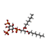
ChemComp-EUJ:
(2R)-3-{[(S)-hydroxy{[(1S,2R,3R,4S,5S,6R)-2,4,6-trihydroxy-3,5-bis(phosphonooxy)cyclohexyl]oxy}phosphoryl]oxy}propane-1,2-diyl dioctanoate

ChemComp-HOH:
WATER

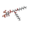
ChemComp-PIO:
[(2R)-2-octanoyloxy-3-[oxidanyl-[(1R,2R,3S,4R,5R,6S)-2,3,6-tris(oxidanyl)-4,5-diphosphonooxy-cyclohexyl]oxy-phosphoryl]oxy-propyl] octanoate

Source
  • homo sapiens (human)
KeywordsLIPID BINDING PROTEIN / ESCRT / ESCRT-III / PIP2 / PI(3 / 5)P2 / endosomal / cytokinesis / PI(4 / CHMP1A

+
About Yorodumi Papers

-
News

-
Feb 9, 2022. New format data for meta-information of EMDB entries

New format data for meta-information of EMDB entries

  • Version 3 of the EMDB header file is now the official format.
  • The previous official version 1.9 will be removed from the archive.

Related info.:EMDB header

External links:wwPDB to switch to version 3 of the EMDB data model

-
Aug 12, 2020. Covid-19 info

Covid-19 info

URL: https://pdbj.org/emnavi/covid19.php

New page: Covid-19 featured information page in EM Navigator.

Related info.:Covid-19 info / Mar 5, 2020. Novel coronavirus structure data

+
Mar 5, 2020. Novel coronavirus structure data

Novel coronavirus structure data

Related info.:Yorodumi Speices / Aug 12, 2020. Covid-19 info

External links:COVID-19 featured content - PDBj / Molecule of the Month (242):Coronavirus Proteases

+
Jan 31, 2019. EMDB accession codes are about to change! (news from PDBe EMDB page)

EMDB accession codes are about to change! (news from PDBe EMDB page)

  • The allocation of 4 digits for EMDB accession codes will soon come to an end. Whilst these codes will remain in use, new EMDB accession codes will include an additional digit and will expand incrementally as the available range of codes is exhausted. The current 4-digit format prefixed with “EMD-” (i.e. EMD-XXXX) will advance to a 5-digit format (i.e. EMD-XXXXX), and so on. It is currently estimated that the 4-digit codes will be depleted around Spring 2019, at which point the 5-digit format will come into force.
  • The EM Navigator/Yorodumi systems omit the EMD- prefix.

Related info.:Q: What is EMD? / ID/Accession-code notation in Yorodumi/EM Navigator

External links:EMDB Accession Codes are Changing Soon! / Contact to PDBj

+
Jul 12, 2017. Major update of PDB

Major update of PDB

  • wwPDB released updated PDB data conforming to the new PDBx/mmCIF dictionary.
  • This is a major update changing the version number from 4 to 5, and with Remediation, in which all the entries are updated.
  • In this update, many items about electron microscopy experimental information are reorganized (e.g. em_software).
  • Now, EM Navigator and Yorodumi are based on the updated data.

External links:wwPDB Remediation / Enriched Model Files Conforming to OneDep Data Standards Now Available in the PDB FTP Archive

-
Yorodumi Papers

Database of articles cited by EMDB/PDB/SASBDB data

  • Database of articles cited by EMDB, PDB, and SASBDB entries
  • Using PubMed data

Related info.:EMDB / PDB / SASBDB / Yorodumi / EMN Papers / Changes in new EM Navigator and Yorodumi

Read more