[English] 日本語
Yorodumi
- EMDB-77060: CHMP1A bound to PI(4,5)P2 containing membrane -

+
Open data


ID or keywords:

Loading...

-
Basic information

Entry
Database: EMDB / ID: EMD-77060
TitleCHMP1A bound to PI(4,5)P2 containing membrane
Map dataSharpenned map
Sample
  • Complex: Charged multivesicular body protein 1a
    • Protein or peptide: Charged multivesicular body protein 1a
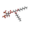
  • Ligand: [(2R)-2-octanoyloxy-3-[oxidanyl-[(1R,2R,3S,4R,5R,6S)-2,3,6-tris(oxidanyl)-4,5-diphosphonooxy-cyclohexyl]oxy-phosphoryl]oxy-propyl] octanoate
  • Ligand: water
KeywordsESCRT / ESCRT-III / PIP2 / PI(4 / 5)P2 / endosomal / cytokinesis / LIPID BINDING PROTEIN / CHMP1A
Function / homology
Function and homology information


multivesicular body-lysosome fusion / amphisome membrane / vesicle fusion with vacuole / ESCRT III complex disassembly / late endosome to lysosome transport / ESCRT III complex / kinetochore microtubule / endosome transport via multivesicular body sorting pathway / nuclear membrane reassembly / multivesicular body sorting pathway ...multivesicular body-lysosome fusion / amphisome membrane / vesicle fusion with vacuole / ESCRT III complex disassembly / late endosome to lysosome transport / ESCRT III complex / kinetochore microtubule / endosome transport via multivesicular body sorting pathway / nuclear membrane reassembly / multivesicular body sorting pathway / midbody abscission / regulation of centrosome duplication / membrane fission / late endosome to vacuole transport / multivesicular body assembly / multivesicular body membrane / ubiquitin-dependent protein catabolic process via the multivesicular body sorting pathway / mitotic chromosome condensation / plasma membrane repair / regulation of mitotic spindle assembly / mitotic metaphase chromosome alignment / nucleus organization / viral budding via host ESCRT complex / microtubule organizing center / autophagosome membrane / autophagosome maturation / nuclear pore / multivesicular body / vesicle-mediated transport / endomembrane system / viral budding from plasma membrane / condensed nuclear chromosome / HCMV Late Events / kinetochore / autophagy / nuclear matrix / metallopeptidase activity / protein transport / midbody / early endosome / protein domain specific binding / negative regulation of gene expression / cell division / lysosomal membrane / DNA-templated transcription / protein homodimerization activity / extracellular exosome / zinc ion binding / nucleoplasm / identical protein binding / plasma membrane / cytosol
Similarity search - Function
Charged multivesicular body protein 1a
Similarity search - Component
Biological speciesHomo sapiens (human)
Methodhelical reconstruction / cryo EM / Resolution: 3.3 Å
AuthorsAlian A / Talledge N / Moss III FR / McCullough J / Frost A / Sundquist WI
Funding support United States, 1 items
OrganizationGrant numberCountry
National Institutes of Health/National Institute Of Allergy and Infectious Diseases (NIH/NIAID)R37 AI051174 United States
CitationJournal: Proc.Natl.Acad.Sci.USA / Year: 2026
Title: Phosphatidylinositol Diphosphate Binding by ESCRT-III Filaments
Authors: Alian A / McCullough J / Moss III FR / Talledge N / Mohammed A / Gerstner C / Dalluge J / Paine E / Davulcu O / Chang CL / Frost A / Sundquist WI
History
DepositionMay 5, 2026-
Header (metadata) releaseJul 8, 2026-
Map releaseJul 8, 2026-
UpdateJul 8, 2026-
Current statusJul 8, 2026Processing site: RCSB / Status: Released

-
Structure visualization

Supplemental images

Downloads & links

-
Map

FileReleased
AnnotationSharpenned map
Projections & slices

Image control

Size
Brightness
Contrast
Others
AxesZ (Sec.)Y (Row.)X (Col.)
1 Å/pix.
x 576 pix.
= 576. Å
1 Å/pix.
x 576 pix.
= 576. Å
1 Å/pix.
x 576 pix.
= 576. Å

Surface

Projections

Slices (1/3)

Slices (1/2)

Slices (2/3)

Images are generated by Spider.

Voxel sizeX=Y=Z: 1 Å
Density
Contour LevelBy AUTHOR: 0.1
Minimum - Maximum-0.4748915 - 1.2137275
Average (Standard dev.)0.012019434 (±0.069338106)
SymmetrySpace group: 1
Details

EMDB XML:

Map geometry
Axis orderXYZ
Origin000
Dimensions576576576
Spacing576576576
CellA=B=C: 576.0 Å
α=β=γ: 90.0 °

-
Supplemental data

-
Mask #1

Fileemd_77060_msk_1.map
Projections & Slices
AxesZYX

Projections

Slices (1/2)
Density Histograms

-
Additional map: Local refinement sharpenned map

Fileemd_77060_additional_1.map
AnnotationLocal refinement sharpenned map
Projections & Slices
AxesZYX

Projections

Slices (1/2)
Density Histograms

-
Additional map: Unsharpenned map

Fileemd_77060_additional_2.map
AnnotationUnsharpenned map
Projections & Slices
AxesZYX

Projections

Slices (1/2)
Density Histograms

-
Half map: Map half-1

Fileemd_77060_half_map_1.map
AnnotationMap half-1
Projections & Slices
AxesZYX

Projections

Slices (1/2)
Density Histograms

-
Half map: Map half-2

Fileemd_77060_half_map_2.map
AnnotationMap half-2
Projections & Slices
AxesZYX

Projections

Slices (1/2)
Density Histograms

-
Sample components

-
Entire : Charged multivesicular body protein 1a

EntireName: Charged multivesicular body protein 1a
Components
  • Complex: Charged multivesicular body protein 1a
    • Protein or peptide: Charged multivesicular body protein 1a
  • Ligand: [(2R)-2-octanoyloxy-3-[oxidanyl-[(1R,2R,3S,4R,5R,6S)-2,3,6-tris(oxidanyl)-4,5-diphosphonooxy-cyclohexyl]oxy-phosphoryl]oxy-propyl] octanoate
  • Ligand: water

-
Supramolecule #1: Charged multivesicular body protein 1a

SupramoleculeName: Charged multivesicular body protein 1a / type: complex / ID: 1 / Parent: 0 / Macromolecule list: #1 / Details: Charged multivesicular body protein 1a
Source (natural)Organism: Homo sapiens (human)
Molecular weightTheoretical: 21.70221 kDa/nm

-
Macromolecule #1: Charged multivesicular body protein 1a

MacromoleculeName: Charged multivesicular body protein 1a / type: protein_or_peptide / ID: 1 / Details: residues 164-196 are unstructured / Number of copies: 1 / Enantiomer: LEVO
Source (natural)Organism: Homo sapiens (human)
Molecular weightTheoretical: 21.731812 KDa
Recombinant expressionOrganism: Escherichia coli 'BL21-Gold(DE3)pLysS AG' (bacteria)
SequenceString: MDDTLFQLKF TAKQLEKLAK KAEKDSKAEQ AKVKKALLQK NVECARVYAE NAIRKKNEGV NWLRMASRVD AVASKVQTAV TMKGVTKNM AQVTKALDKA LSTMDLQKVS SVMDRFEQQV QNLDVHTSVM EDSMSSATTL TTPQEQVDSL IMQIAEENGL E VLDQLSQL ...String:
MDDTLFQLKF TAKQLEKLAK KAEKDSKAEQ AKVKKALLQK NVECARVYAE NAIRKKNEGV NWLRMASRVD AVASKVQTAV TMKGVTKNM AQVTKALDKA LSTMDLQKVS SVMDRFEQQV QNLDVHTSVM EDSMSSATTL TTPQEQVDSL IMQIAEENGL E VLDQLSQL PEGASAVGES SVRSQEDQLS RRLAALRN

UniProtKB: Charged multivesicular body protein 1a

-
Macromolecule #2: [(2R)-2-octanoyloxy-3-[oxidanyl-[(1R,2R,3S,4R,5R,6S)-2,3,6-tris(o...

MacromoleculeName: [(2R)-2-octanoyloxy-3-[oxidanyl-[(1R,2R,3S,4R,5R,6S)-2,3,6-tris(oxidanyl)-4,5-diphosphonooxy-cyclohexyl]oxy-phosphoryl]oxy-propyl] octanoate
type: ligand / ID: 2 / Number of copies: 1 / Formula: PIO
Molecular weightTheoretical: 746.566 Da
Chemical component information

ChemComp-PIO:
[(2R)-2-octanoyloxy-3-[oxidanyl-[(1R,2R,3S,4R,5R,6S)-2,3,6-tris(oxidanyl)-4,5-diphosphonooxy-cyclohexyl]oxy-phosphoryl]oxy-propyl] octanoate

-
Macromolecule #3: water

MacromoleculeName: water / type: ligand / ID: 3 / Number of copies: 1 / Formula: HOH
Molecular weightTheoretical: 18.015 Da
Chemical component information

ChemComp-HOH:
WATER

-
Experimental details

-
Structure determination

Methodcryo EM
Processinghelical reconstruction
Aggregation statefilament

-
Sample preparation

BufferpH: 8
VitrificationCryogen name: ETHANE / Chamber humidity: 85 % / Chamber temperature: 284 K / Instrument: LEICA EM GP

-
Electron microscopy

MicroscopeTFS KRIOS
Image recordingFilm or detector model: GATAN K3 (6k x 4k) / Number grids imaged: 2 / Number real images: 73224 / Average electron dose: 46.0 e/Å2
Electron beamAcceleration voltage: 300 kV / Electron source: FIELD EMISSION GUN
Electron opticsC2 aperture diameter: 50.0 µm / Illumination mode: FLOOD BEAM / Imaging mode: OTHER / Nominal defocus max: 2.2 µm / Nominal defocus min: 0.8 µm
Experimental equipment
Model: Titan Krios / Image courtesy: FEI Company

+
Image processing

DetailsAll images were processed using the same pipeline
Final reconstructionApplied symmetry - Helical parameters - Δz: 1.19 Å
Applied symmetry - Helical parameters - Δ&Phi: 10.948 °
Applied symmetry - Helical parameters - Axial symmetry: C1 (asymmetric)
Resolution.type: BY AUTHOR / Resolution: 3.3 Å / Resolution method: FSC 0.143 CUT-OFF / Software - Name: cryoSPARC (ver. 5.0.4) / Number images used: 150055
CTF correctionType: NONE
Segment selectionNumber selected: 1050919 / Software - Name: RELION (ver. 5.0-beta)
Startup modelType of model: INSILICO MODEL / In silico model: ModelAngelo and AlphaFold
Final angle assignmentType: NOT APPLICABLE
FSC plot (resolution estimation)

-
Atomic model buiding 1

Initial model
ChainDetails
source_name: Other, initial_model_type: in silico modelModelAngelo
source_name: AlphaFold, initial_model_type: in silico model
RefinementSpace: REAL / Protocol: AB INITIO MODEL / Overall B value: 107.8
Output model

PDB-13gm:
CHMP1A bound to PI(4,5)P2 containing membrane

+
About Yorodumi

-
News

-
Feb 9, 2022. New format data for meta-information of EMDB entries

New format data for meta-information of EMDB entries

  • Version 3 of the EMDB header file is now the official format.
  • The previous official version 1.9 will be removed from the archive.

Related info.:EMDB header

External links:wwPDB to switch to version 3 of the EMDB data model

-
Aug 12, 2020. Covid-19 info

Covid-19 info

URL: https://pdbj.org/emnavi/covid19.php

New page: Covid-19 featured information page in EM Navigator.

Related info.:Covid-19 info / Mar 5, 2020. Novel coronavirus structure data

+
Mar 5, 2020. Novel coronavirus structure data

Novel coronavirus structure data

Related info.:Yorodumi Speices / Aug 12, 2020. Covid-19 info

External links:COVID-19 featured content - PDBj / Molecule of the Month (242):Coronavirus Proteases

+
Jan 31, 2019. EMDB accession codes are about to change! (news from PDBe EMDB page)

EMDB accession codes are about to change! (news from PDBe EMDB page)

  • The allocation of 4 digits for EMDB accession codes will soon come to an end. Whilst these codes will remain in use, new EMDB accession codes will include an additional digit and will expand incrementally as the available range of codes is exhausted. The current 4-digit format prefixed with “EMD-” (i.e. EMD-XXXX) will advance to a 5-digit format (i.e. EMD-XXXXX), and so on. It is currently estimated that the 4-digit codes will be depleted around Spring 2019, at which point the 5-digit format will come into force.
  • The EM Navigator/Yorodumi systems omit the EMD- prefix.

Related info.:Q: What is EMD? / ID/Accession-code notation in Yorodumi/EM Navigator

External links:EMDB Accession Codes are Changing Soon! / Contact to PDBj

+
Jul 12, 2017. Major update of PDB

Major update of PDB

  • wwPDB released updated PDB data conforming to the new PDBx/mmCIF dictionary.
  • This is a major update changing the version number from 4 to 5, and with Remediation, in which all the entries are updated.
  • In this update, many items about electron microscopy experimental information are reorganized (e.g. em_software).
  • Now, EM Navigator and Yorodumi are based on the updated data.

External links:wwPDB Remediation / Enriched Model Files Conforming to OneDep Data Standards Now Available in the PDB FTP Archive

-
Yorodumi

Thousand views of thousand structures

  • Yorodumi is a browser for structure data from EMDB, PDB, SASBDB, etc.
  • This page is also the successor to EM Navigator detail page, and also detail information page/front-end page for Omokage search.
  • The word "yorodu" (or yorozu) is an old Japanese word meaning "ten thousand". "mi" (miru) is to see.

Related info.:EMDB / PDB / SASBDB / Comparison of 3 databanks / Yorodumi Search / Aug 31, 2016. New EM Navigator & Yorodumi / Yorodumi Papers / Jmol/JSmol / Function and homology information / Changes in new EM Navigator and Yorodumi

Read more