 Movie
Movie Controller


+ Open data

- Basic information
| Entry | Database: PDB / ID: 13gm | |||||||||||||||||||||||||||||||||
|---|---|---|---|---|---|---|---|---|---|---|---|---|---|---|---|---|---|---|---|---|---|---|---|---|---|---|---|---|---|---|---|---|---|---|
| Title | CHMP1A bound to PI(4,5)P2 containing membrane | |||||||||||||||||||||||||||||||||
 Components Components | Charged multivesicular body protein 1a | |||||||||||||||||||||||||||||||||
 Keywords Keywords | LIPID BINDING PROTEIN / ESCRT / ESCRT-III / PIP2 / PI(4 / 5)P2 / endosomal / cytokinesis / CHMP1A | |||||||||||||||||||||||||||||||||
| Function / homology |  Function and homology information Function and homology informationmultivesicular body-lysosome fusion / amphisome membrane / vesicle fusion with vacuole / ESCRT III complex disassembly / late endosome to lysosome transport / ESCRT III complex / kinetochore microtubule / nuclear membrane reassembly / multivesicular body sorting pathway / midbody abscission ...multivesicular body-lysosome fusion / amphisome membrane / vesicle fusion with vacuole / ESCRT III complex disassembly / late endosome to lysosome transport / ESCRT III complex / kinetochore microtubule / nuclear membrane reassembly / multivesicular body sorting pathway / midbody abscission / membrane fission / plasma membrane repair / multivesicular body membrane / multivesicular body assembly / ubiquitin-dependent protein catabolic process via the multivesicular body sorting pathway / nucleus organization / late endosome to vacuole transport / mitotic chromosome condensation / regulation of mitotic spindle assembly / regulation of centrosome duplication / viral budding via host ESCRT complex / microtubule organizing center / autophagosome membrane / autophagosome maturation / mitotic metaphase chromosome alignment / nuclear pore / vesicle-mediated transport / viral budding from plasma membrane / condensed nuclear chromosome / HCMV Late Events / endomembrane system / autophagy / kinetochore / metallopeptidase activity / nuclear matrix / cell division / midbody / early endosome / negative regulation of gene expression / protein domain specific binding / lysosomal membrane / protein homodimerization activity / extracellular exosome / zinc ion binding / nucleoplasm / identical protein binding / plasma membrane / cytosol Similarity search - Function | |||||||||||||||||||||||||||||||||
| Biological species |  Homo sapiens (human) Homo sapiens (human) | |||||||||||||||||||||||||||||||||
| Method | ELECTRON MICROSCOPY / helical reconstruction / cryo EM / Resolution: 3.3 Å | |||||||||||||||||||||||||||||||||
 Authors Authors | Alian, A. / Talledge, N. / Moss III, F.R. / McCullough, J. / Frost, A. / Sundquist, W.I. | |||||||||||||||||||||||||||||||||
| Funding support |  United States, 1items United States, 1items
| |||||||||||||||||||||||||||||||||
 Citation Citation | Journal: Proc Natl Acad Sci U S A / Year: 2026 Title: Phosphatidylinositol diphosphate binding by ESCRT-III filaments. Authors: Akram Alian / John McCullough / Frank R Moss / Nathaniel Talledge / Arshad Mohammed / Cecilia D Gerstner / Jacob A Dalluge / Elliott L Paine / Omar Davulcu / Chi-Lun Chang / Adam Frost / Wesley I Sundquist / Abstract: Different inositol phospholipids (PIPs) distribute to distinct subcellular organelles, creating an addressing system that dictates the sites of action of PIP-binding proteins, including components of ...Different inositol phospholipids (PIPs) distribute to distinct subcellular organelles, creating an addressing system that dictates the sites of action of PIP-binding proteins, including components of the Endosomal Sorting Complexes Required for Transport (ESCRT). The ESCRT machinery is recruited to remodel many different cellular membranes through combinatorial binding interactions made by the early-acting ESCRT-I and ESCRT-II complexes with PIPs, ubiquitin modifications, and membrane-specific adaptors. Membrane remodeling, constriction, and fission are then mediated by membrane-associated filaments formed by subunits of the late-acting ESCRT-III complexes, together with their associated VPS4 AAA ATPases. Here, we describe two different classes of helical ESCRT-III filaments that can surround and tubulate membranes containing PIP lipids. Cryo-EM reconstructions revealed that protofilaments comprising closed IST1 subunits formed 8-stranded nanotubes that encase membrane monolayers. The nanotube coordinates exposed PI(4,5)P or PI(3,5)P headgroups within a basic pocket formed at the junction of three IST1 subunits, and our structures reveal how the pocket can accommodate either PIP isomer with minimal adjustment. In contrast, protofilaments comprising open CHMP1A subunits formed one start helices that encase membrane bilayers and bind exposed PI(4,5)P headgroups across a basic surface that spans adjacent subunits of the CHMP1A protofilament. These two different structures extend the known plasticity of ESCRT-III polymers, reveal how PIP lipids can promote ESCRT-III filament assembly and membrane remodeling, and define the molecular contacts that underlie specific ESCRT-III/PIP interactions. | |||||||||||||||||||||||||||||||||
| History |
|
- Structure visualization
| Structure viewer | Molecule: MolmilJmol/JSmol |
|---|
- Downloads & links
-Download
| PDBx/mmCIF format | 13gm.cif.gz | 69.2 KB | Display | PDBx/mmCIF format |
|---|---|---|---|---|
| PDB format | pdb13gm.ent.gz | 50.5 KB | Display | PDB format |
| PDBx/mmJSON format | 13gm.json.gz | Tree view | PDBx/mmJSON format | |
| Others |  Other downloads Other downloads |
-Validation report
| Arichive directory | https://data.pdbj.org/pub/pdb/validation_reports/3g/13gmftp://data.pdbj.org/pub/pdb/validation_reports/3g/13gm | HTTPS FTP |
|---|
-Related structure data
| Related structure data |  77060MC  13ghC  13gjC C: citing same article ( M: map data used to model this data |
|---|---|
| Similar structure data |


-Links
 PDBj
PDBj

- Assembly
| Deposited unit | 
|
|---|---|
| 1 | x 81
|
-Components
| #1: Protein | Mass: 21731.812 Da / Num. of mol.: 1 Source method: isolated from a genetically manipulated source Details: residues 164-196 are unstructured / Source: (gene. exp.) Homo sapiens (human) / Gene: CHMP1A, CHMP1, KIAA0047, PCOLN3, PRSM1Production host:   Escherichia coli 'BL21-Gold(DE3)pLysS AG' (bacteria) Escherichia coli 'BL21-Gold(DE3)pLysS AG' (bacteria)References: UniProt: Q9HD42 |
|---|---|
| #2: Chemical | ChemComp-PIO / [(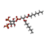  Mass: 746.566 Da / Num. of mol.: 1 / Source method: obtained synthetically / Formula: C25H49O19P3 / Feature type: SUBJECT OF INVESTIGATION Mass: 746.566 Da / Num. of mol.: 1 / Source method: obtained synthetically / Formula: C25H49O19P3 / Feature type: SUBJECT OF INVESTIGATION |
| #3: Water | ChemComp-HOH /  Mass: 18.015 Da / Num. of mol.: 1 / Source method: isolated from a natural source / Formula: H2O Mass: 18.015 Da / Num. of mol.: 1 / Source method: isolated from a natural source / Formula: H2O |
| Has ligand of interest | Y |
| Has protein modification | N |
-Experimental details
-Experiment
| Experiment | Method: ELECTRON MICROSCOPY |
|---|---|
| EM experiment | Aggregation state: FILAMENT / 3D reconstruction method: helical reconstruction |
- Sample preparation
| Component | Name: Charged multivesicular body protein 1a / Type: COMPLEX / Details: Charged multivesicular body protein 1a / Entity ID: #1 / Source: RECOMBINANT |
|---|---|
| Molecular weight | Value: 21.70221 kDa/nm / Experimental value: YES |
| Source (natural) | Organism: Homo sapiens (human) |
| Source (recombinant) | Organism: Escherichia coli 'BL21-Gold(DE3)pLysS AG' (bacteria) |
| Buffer solution | pH: 8 |
| Specimen | Embedding applied: NO / Shadowing applied: NO / Staining applied: NO / Vitrification applied: YES |
| Vitrification | Instrument: LEICA EM GP / Cryogen name: ETHANE / Humidity: 85 % / Chamber temperature: 284 K |
- Electron microscopy imaging
| Experimental equipment |  Model: Titan Krios / Image courtesy: FEI Company |
|---|---|
| Microscopy | Model: TFS KRIOS |
| Electron gun | Electron source:  FIELD EMISSION GUN / Accelerating voltage: 300 kV / Illumination mode: FLOOD BEAM FIELD EMISSION GUN / Accelerating voltage: 300 kV / Illumination mode: FLOOD BEAM |
| Electron lens | Mode: OTHER / Nominal defocus max: 2200 nm / Nominal defocus min: 800 nm / C2 aperture diameter: 50 µm |
| Image recording | Electron dose: 46 e/Å2 / Film or detector model: GATAN K3 (6k x 4k) / Num. of grids imaged: 2 / Num. of real images: 73224 |
- Processing
| EM software |
| ||||||||||||||||||||||||||||
|---|---|---|---|---|---|---|---|---|---|---|---|---|---|---|---|---|---|---|---|---|---|---|---|---|---|---|---|---|---|
| Image processing | Details: All images were processed using the same pipeline | ||||||||||||||||||||||||||||
| CTF correction | Type: NONE | ||||||||||||||||||||||||||||
| Helical symmerty | Angular rotation/subunit: 10.948 ° / Axial rise/subunit: 1.19 Å / Axial symmetry: C1 | ||||||||||||||||||||||||||||
| Particle selection | Num. of particles selected: 1050919 | ||||||||||||||||||||||||||||
| 3D reconstruction | Resolution: 3.3 Å / Resolution method: FSC 0.143 CUT-OFF / Num. of particles: 150055 / Symmetry type: HELICAL | ||||||||||||||||||||||||||||
| Atomic model building | B value: 107.8 / Protocol: AB INITIO MODEL / Space: REAL | ||||||||||||||||||||||||||||
| Atomic model building |
|
