 Movie
Movie Controller
 Structure viewers
 About Yorodumi Papers


+Search query
-Structure paper
| Title | Structural characterisation of the fungal Pmt4 homodimer. |
|---|---|
| Journal, issue, pages | Nat Commun, Vol. 16, Issue 1, Page 11134, Year 2025 |
| Publish date | Dec 14, 2025 |
 Authors Authors | Melanie A McDowell / Klemens Wild / Francesco Fiorentino / Daniela Bausewein / Anke Metschies / Antonella Chiapparino / Yvonne Hackmann / Florestan L Bilsing / David Brenske / Sofia Mortensen / Di Wu / Carol V Robinson / Sabine Strahl / Irmgard Sinning /    |
| PubMed Abstract | Protein O-mannosyltransferases (PMTs) are conserved endoplasmic reticulum membrane-embedded enzymes responsible for the transfer of mannose from dolichol phosphate-mannose (Dol-P-Man) to ...Protein O-mannosyltransferases (PMTs) are conserved endoplasmic reticulum membrane-embedded enzymes responsible for the transfer of mannose from dolichol phosphate-mannose (Dol-P-Man) to serine/threonine-rich protein substrates or unfolded proteins. PMTs from three subfamilies form obligate dimers with different substrate specificities and require the concerted action of their transmembrane domains (TMDs) and a luminal MIR domain for catalysis. Here, we present structures, native mass spectrometry, and structure-based mutagenesis of the fungal Pmt4 homodimer. The core fold of the TMDs and MIR domain is conserved with the Pmt1-Pmt2 heterodimer, indicating a shared catalytic mechanism. Distinct from Pmt4, the MIR domain interacts in cis with the TMDs of the same subunit and has a β-hairpin insertion required for O-mannosylation of substrates. We further identify a cytosolic binding site for substrate Dol-P-Man within the Pmt4 TMDs, which is conserved amongst PMTs and important for in vivo activity. Thus, we provide a framework to understand the substrate specificity and regulation of the Pmt4 homodimer. |
 External links External links | Nat Commun / PubMed:41392315 / PubMed Central |
| Methods | EM (single particle) / X-ray diffraction |
| Resolution | 1.22 - 3.4 Å |
| Structure data | EMDB-52631, PDB-9i5k: EMDB-52632, PDB-9i5l:  PDB-9fd0:  PDB-9fd1: |
| Chemicals |  ChemComp-EPE:  ChemComp-GOL:  ChemComp-HOH:  ChemComp-EDO:  ChemComp-ACT:  ChemComp-NA: 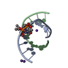 PDB-1i0n: 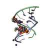 PDB-1i0m: |
| Source |
|
 Keywords Keywords | PEPTIDE BINDING PROTEIN / Protein O-mannosylation / glycosylation / ER luminal domain / b-trefoil / ligand binding / processivity / thermostability / MEMBRANE PROTEIN / homodimer / ER / biogenesis |


 thermochaetoides thermophila (fungus)
