 Movie
Movie Controller
 Structure viewers
 About Yorodumi Papers


+Search query
-Structure paper
| Title | Helical superstructures between amyloid and collagen in cardiac fibrils from a patient with AL amyloidosis. |
|---|---|
| Journal, issue, pages | Nat Commun, Vol. 15, Issue 1, Page 6359, Year 2024 |
| Publish date | Jul 28, 2024 |
 Authors Authors | Tim Schulte / Antonio Chaves-Sanjuan / Valentina Speranzini / Kevin Sicking / Melissa Milazzo / Giulia Mazzini / Paola Rognoni / Serena Caminito / Paolo Milani / Chiara Marabelli / Alessandro Corbelli / Luisa Diomede / Fabio Fiordaliso / Luigi Anastasia / Carlo Pappone / Giampaolo Merlini / Martino Bolognesi / Mario Nuvolone / Rubén Fernández-Busnadiego / Giovanni Palladini / Stefano Ricagno /     |
| PubMed Abstract | Systemic light chain (LC) amyloidosis (AL) is a disease where organs are damaged by an overload of a misfolded patient-specific antibody-derived LC, secreted by an abnormal B cell clone. The high LC ...Systemic light chain (LC) amyloidosis (AL) is a disease where organs are damaged by an overload of a misfolded patient-specific antibody-derived LC, secreted by an abnormal B cell clone. The high LC concentration in the blood leads to amyloid deposition at organ sites. Indeed, cryogenic electron microscopy (cryo-EM) has revealed unique amyloid folds for heart-derived fibrils taken from different patients. Here, we present the cryo-EM structure of heart-derived AL amyloid (AL59) from another patient with severe cardiac involvement. The double-layered structure displays a u-shaped core that is closed by a β-arc lid and extended by a straight tail. Noteworthy, the fibril harbours an extended constant domain fragment, thus ruling out the variable domain as sole amyloid building block. Surprisingly, the fibrils were abundantly concatenated with a proteinaceous polymer, here identified as collagen VI (COLVI) by immuno-electron microscopy (IEM) and mass-spectrometry. Cryogenic electron tomography (cryo-ET) showed how COLVI wraps around the amyloid forming a helical superstructure, likely stabilizing and protecting the fibrils from clearance. Thus, here we report structural evidence of interactions between amyloid and collagen, potentially signifying a distinct pathophysiological mechanism of amyloid deposits. |
 External links External links | Nat Commun / PubMed:39069558 / PubMed Central |
| Methods | EM (single particle) / EM (helical sym.) / EM (tomography) |
| Resolution | 3.6 - 15.0 Å |
| Structure data | 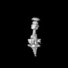 EMDB-18689: Maps of Collagen VI half- and full-beads EMDB-50270, PDB-9faa: EMDB-50272, PDB-9fac:  EMDB-51031: Cryo-electron tomogram of AL59 amyloids interacting with collagen VI  EMDB-51032: Cryo-electron tomogram of AL59 amyloids interacting with collagen VI  EMDB-51033: Cryo-electron tomogram of AL59 amyloids interacting with collagen VI  EMDB-51038: Cryo-electron tomogram of AL59 amyloids interacting with collagen VI |
| Chemicals |  ChemComp-NAG: |
| Source |
|
 Keywords Keywords | PROTEIN FIBRIL / systemic AL amyloid fibril |


 homo sapiens (human)