 Movie
Movie Controller
 Structure viewers
 About Yorodumi Papers


+Search query
-Structure paper
| Title | Cryo-EM structures of a protein pore reveal a cluster of cholesterol molecules and diverse roles of membrane lipids. |
|---|---|
| Journal, issue, pages | Nat Commun, Vol. 16, Issue 1, Page 2972, Year 2025 |
| Publish date | Mar 26, 2025 |
 Authors Authors | Gašper Šolinc / Marija Srnko / Franci Merzel / Ana Crnković / Mirijam Kozorog / Marjetka Podobnik / Gregor Anderluh /  |
| PubMed Abstract | The structure and function of membrane proteins depend on their interactions with lipids that constitute membranes. Actinoporins are α-pore-forming proteins that bind preferentially to sphingomyelin- ...The structure and function of membrane proteins depend on their interactions with lipids that constitute membranes. Actinoporins are α-pore-forming proteins that bind preferentially to sphingomyelin-containing membranes, where they oligomerize and form transmembrane pores. Through a comprehensive cryo-electron microscopic analysis of a pore formed by an actinoporin Fav from the coral Orbicella faveolata, we show that the octameric pore interacts with 112 lipids in the upper leaflet of the membrane, reveal the roles of lipids, and demonstrate that the actinoporin surface is suited for binding multiple receptor sphingomyelin molecules. When cholesterol is present in the membrane, it forms a cluster of four molecules associated with each protomer. Atomistic simulations support the structural data and reveal additional effects of the pore on the lipid membrane. These data reveal a complex network of protein-lipid and lipid-lipid interactions and an underrated role of lipids in the structure and function of transmembrane protein complexes. |
 External links External links | Nat Commun / PubMed:40140423 / PubMed Central |
| Methods | EM (single particle) / X-ray diffraction |
| Resolution | 1.5 - 3.6 Å |
| Structure data | EMDB-50057, PDB-9eym: EMDB-50058, PDB-9eyn: EMDB-50059, PDB-9eyo:  EMDB-50060: The octameric pore of actinoporin Fav prepared on DOPC:sphingomyelin:cholesterol containing nanodiscs  PDB-9eyl: |
| Chemicals |  ChemComp-SO4: 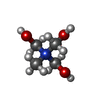 ChemComp-144:  ChemComp-HOH: 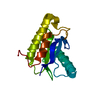 PDB-1h8m:  ChemComp-CLR: |
| Source |
|
 Keywords Keywords | TOXIN / Actinoporin / Pore-forming toxin / Orbicella faveolata / Pore / Octamer / Transmembrane pore / Nanopore / cholesterol |


 orbicella faveolata (invertebrata)