[English] 日本語
Yorodumi Papers
- Database of articles cited by EMDB/PDB/SASBDB data -

+
Search query

Keywords
Structure methods
Author
Journal
IF

-
Structure paper

TitleMolecular basis of global promoter sensing and nucleosome capture by the SWR1 chromatin remodeler.
Journal, issue, pagesCell, Vol. 187, Issue 24, Page 6849-6864.e18, Year 2024
Publish dateNov 27, 2024
AuthorsRobert K Louder / Giho Park / Ziyang Ye / Justin S Cha / Anne M Gardner / Qin Lei / Anand Ranjan / Eva Höllmüller / Florian Stengel / B Franklin Pugh / Carl Wu /
PubMed AbstractThe SWR1 chromatin remodeling complex is recruited to +1 nucleosomes downstream of transcription start sites of eukaryotic promoters, where it exchanges histone H2A for the specialized variant H2A.Z. ...The SWR1 chromatin remodeling complex is recruited to +1 nucleosomes downstream of transcription start sites of eukaryotic promoters, where it exchanges histone H2A for the specialized variant H2A.Z. Here, we use cryoelectron microscopy (cryo-EM) to resolve the structural basis of the SWR1 interaction with free DNA, revealing a distinct open conformation of the Swr1 ATPase that enables sliding from accessible DNA to nucleosomes. A complete structural model of the SWR1-nucleosome complex illustrates critical roles for Swc2 and Swc3 subunits in oriented nucleosome engagement by SWR1. Moreover, an extended DNA-binding α helix within the Swc3 subunit enables sensing of nucleosome linker length and is essential for SWR1-promoter-specific recruitment and activity. The previously unresolved N-SWR1 subcomplex forms a flexible extended structure, enabling multivalent recognition of acetylated histone tails by reader domains to further direct SWR1 toward the +1 nucleosome. Altogether, our findings provide a generalizable mechanism for promoter-specific targeting of chromatin and transcription complexes.
External linksCell / PubMed:39357520 / PubMed Central
MethodsEM (single particle)
Resolution3.3 - 7.3 Å
Structure data

EMDB-44074, PDB-9b1d:
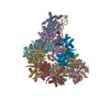
Cryo-EM structure of native SWR1 bound to DNA (composite structure)
Method: EM (single particle) / Resolution: 3.3 Å

EMDB-44075, PDB-9b1e:
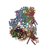
Cryo-EM structure of native SWR1 bound to nucleosome (composite structure)
Method: EM (single particle) / Resolution: 4.4 Å

EMDB-44093: Cryo-EM structure of native SWR1, free complex (composite structure)
Method: EM (single particle) / Resolution: 3.9 Å

EMDB-44106: Cryo-EM structure of native SWR1 bound to DNA (consensus map)
Method: EM (single particle) / Resolution: 3.7 Å

EMDB-44107: RuvBL core from SWR1-DNA complex (focused refinement)
Method: EM (single particle) / Resolution: 3.3 Å

EMDB-44108: Swr1 ATPase domain from SWR1-DNA complex (focused refinement)
Method: EM (single particle) / Resolution: 4.5 Å

EMDB-44109: Arp6/Swc6 module from SWR1-DNA complex (focused refinement)
Method: EM (single particle) / Resolution: 3.6 Å

EMDB-44110: Cryo-EM structure of native SWR1 bound to DNA (unmasked refinement filtered by local resolution)
Method: EM (single particle) / Resolution: 3.4 Å

EMDB-44307: Cryo-EM structure of native SWR1 bound to nucleosome (consensus map filtered by local resolution)
Method: EM (single particle) / Resolution: 6.2 Å

EMDB-44308: RuvBL-associated core from SWR1-nucleosome complex (focused refinement)
Method: EM (single particle) / Resolution: 4.4 Å

EMDB-44309: Nucleosome and bound Swr1 ATPase from SWR1-nucleosome complex (focused refinement)
Method: EM (single particle) / Resolution: 4.4 Å

EMDB-44310: Swc3-Swc2 subcomplex from SWR1-nucleosome complex (focused refinement)
Method: EM (single particle) / Resolution: 7.3 Å

EMDB-44311: Cryo-EM structure of native SWR1, free complex (consensus map filtered by local resolution)
Method: EM (single particle) / Resolution: 4.8 Å

EMDB-44312: RuvBL core from free SWR1 complex (focused refinement)
Method: EM (single particle) / Resolution: 3.9 Å

EMDB-44313: Arp6/Swc6 module from free SWR1 complex (focused refinement)
Method: EM (single particle) / Resolution: 4.4 Å

EMDB-46065: Cryo-EM structure of native SWR1 bound to DNA in the absence of nucleotide (composite structure)
Method: EM (single particle) / Resolution: 5.8 Å

EMDB-46066: Cryo-EM structure of native SWR1 bound to DNA in the absence of nucleotide (consensus map)
Method: EM (single particle) / Resolution: 5.8 Å

EMDB-46067: RuvBL core from SWR1(apo)-DNA complex (focused refinement)
Method: EM (single particle) / Resolution: 5.5 Å

EMDB-46068: Arp6/Swc6 module from SWR1(apo)-DNA complex (focused refinement)
Method: EM (single particle) / Resolution: 7.2 Å

EMDB-46069: Swr1 ATPase domain from SWR1(apo)-DNA complex (focused refinement)
Method: EM (single particle) / Resolution: 7.1 Å

Chemicals

ChemComp-AGS:
PHOSPHOTHIOPHOSPHORIC ACID-ADENYLATE ESTER / ATP-gamma-S, energy-carrying molecule analogue*YM

ChemComp-MG:
Unknown entry

ChemComp-ZN:
Unknown entry

ChemComp-ADP:
ADENOSINE-5'-DIPHOSPHATE / ADP, energy-carrying molecule*YM

ChemComp-BEF:
BERYLLIUM TRIFLUORIDE ION

Source
  • saccharomyces cerevisiae (brewer's yeast)
  • saccharomyces cerevisiae w303 (yeast)
  • synthetic construct (others)
  • drosophila melanogaster (fruit fly)
KeywordsGENE REGULATION / Chromatin Remodeler / Snf2 family ATPase / histone exchange / H2A.Z

+
About Yorodumi Papers

-
News

-
Feb 9, 2022. New format data for meta-information of EMDB entries

New format data for meta-information of EMDB entries

  • Version 3 of the EMDB header file is now the official format.
  • The previous official version 1.9 will be removed from the archive.

Related info.:EMDB header

External links:wwPDB to switch to version 3 of the EMDB data model

-
Aug 12, 2020. Covid-19 info

Covid-19 info

URL: https://pdbj.org/emnavi/covid19.php

New page: Covid-19 featured information page in EM Navigator.

Related info.:Covid-19 info / Mar 5, 2020. Novel coronavirus structure data

+
Mar 5, 2020. Novel coronavirus structure data

Novel coronavirus structure data

Related info.:Yorodumi Speices / Aug 12, 2020. Covid-19 info

External links:COVID-19 featured content - PDBj / Molecule of the Month (242):Coronavirus Proteases

+
Jan 31, 2019. EMDB accession codes are about to change! (news from PDBe EMDB page)

EMDB accession codes are about to change! (news from PDBe EMDB page)

  • The allocation of 4 digits for EMDB accession codes will soon come to an end. Whilst these codes will remain in use, new EMDB accession codes will include an additional digit and will expand incrementally as the available range of codes is exhausted. The current 4-digit format prefixed with “EMD-” (i.e. EMD-XXXX) will advance to a 5-digit format (i.e. EMD-XXXXX), and so on. It is currently estimated that the 4-digit codes will be depleted around Spring 2019, at which point the 5-digit format will come into force.
  • The EM Navigator/Yorodumi systems omit the EMD- prefix.

Related info.:Q: What is EMD? / ID/Accession-code notation in Yorodumi/EM Navigator

External links:EMDB Accession Codes are Changing Soon! / Contact to PDBj

+
Jul 12, 2017. Major update of PDB

Major update of PDB

  • wwPDB released updated PDB data conforming to the new PDBx/mmCIF dictionary.
  • This is a major update changing the version number from 4 to 5, and with Remediation, in which all the entries are updated.
  • In this update, many items about electron microscopy experimental information are reorganized (e.g. em_software).
  • Now, EM Navigator and Yorodumi are based on the updated data.

External links:wwPDB Remediation / Enriched Model Files Conforming to OneDep Data Standards Now Available in the PDB FTP Archive

-
Yorodumi Papers

Database of articles cited by EMDB/PDB/SASBDB data

  • Database of articles cited by EMDB, PDB, and SASBDB entries
  • Using PubMed data

Related info.:EMDB / PDB / SASBDB / Yorodumi / EMN Papers / Changes in new EM Navigator and Yorodumi

Read more