[English] 日本語
 Yorodumi
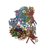
- PDB-9b1d: Cryo-EM structure of native SWR1 bound to DNA (composite structure) -
+ Open data
- Basic information
| Entry | Database: PDB / ID: 9b1d | ||||||||||||
|---|---|---|---|---|---|---|---|---|---|---|---|---|---|
| Title | Cryo-EM structure of native SWR1 bound to DNA (composite structure) | ||||||||||||
 Components Components |
| ||||||||||||
 Keywords Keywords | GENE REGULATION / Chromatin Remodeler / Snf2 family ATPase / histone exchange / H2A.Z | ||||||||||||
| Function / homology |  Function and homology information Function and homology informationTTT Hsp90 cochaperone complex / R2TP complex / protein targeting to vacuole / Swr1 complex / Ino80 complex / box C/D snoRNP assembly / 3'-5' DNA helicase activity / NuA4 histone acetyltransferase complex / nucleosome binding / DNA helicase activity ...TTT Hsp90 cochaperone complex / R2TP complex / protein targeting to vacuole / Swr1 complex / Ino80 complex / box C/D snoRNP assembly / 3'-5' DNA helicase activity / NuA4 histone acetyltransferase complex / nucleosome binding / DNA helicase activity / nuclear periphery / rRNA processing / 5'-3' DNA helicase activity / histone binding / DNA helicase / protein stabilization / chromatin remodeling / DNA repair / regulation of DNA-templated transcription / regulation of transcription by RNA polymerase II / chromatin / ATP hydrolysis activity / zinc ion binding / ATP binding / nucleus / cytoplasm Similarity search - Function | ||||||||||||
| Biological species |  synthetic construct (others) | ||||||||||||
| Method | ELECTRON MICROSCOPY / single particle reconstruction / cryo EM / Resolution: 3.3 Å | ||||||||||||
 Authors Authors | Louder, R.K. / Park, G. / Wu, C. | ||||||||||||
| Funding support |  United States, 3items United States, 3items
| ||||||||||||
 Citation Citation |  Journal: Cell / Year: 2024 Journal: Cell / Year: 2024Title: Molecular basis of global promoter sensing and nucleosome capture by the SWR1 chromatin remodeler. Authors: Robert K Louder / Giho Park / Ziyang Ye / Justin S Cha / Anne M Gardner / Qin Lei / Anand Ranjan / Eva Höllmüller / Florian Stengel / B Franklin Pugh / Carl Wu /   Abstract: The SWR1 chromatin remodeling complex is recruited to +1 nucleosomes downstream of transcription start sites of eukaryotic promoters, where it exchanges histone H2A for the specialized variant H2A.Z. ...The SWR1 chromatin remodeling complex is recruited to +1 nucleosomes downstream of transcription start sites of eukaryotic promoters, where it exchanges histone H2A for the specialized variant H2A.Z. Here, we use cryoelectron microscopy (cryo-EM) to resolve the structural basis of the SWR1 interaction with free DNA, revealing a distinct open conformation of the Swr1 ATPase that enables sliding from accessible DNA to nucleosomes. A complete structural model of the SWR1-nucleosome complex illustrates critical roles for Swc2 and Swc3 subunits in oriented nucleosome engagement by SWR1. Moreover, an extended DNA-binding α helix within the Swc3 subunit enables sensing of nucleosome linker length and is essential for SWR1-promoter-specific recruitment and activity. The previously unresolved N-SWR1 subcomplex forms a flexible extended structure, enabling multivalent recognition of acetylated histone tails by reader domains to further direct SWR1 toward the +1 nucleosome. Altogether, our findings provide a generalizable mechanism for promoter-specific targeting of chromatin and transcription complexes. | ||||||||||||
| History |
|
- Structure visualization
| Structure viewer | Molecule:  Molmil Molmil Jmol/JSmol Jmol/JSmol |
|---|
- Downloads & links
- Download
| PDBx/mmCIF format |  9b1d.cif.gz 9b1d.cif.gz | 1 MB | Display |  PDBx/mmCIF format PDBx/mmCIF format |
|---|---|---|---|---|
| PDB format |  pdb9b1d.ent.gz pdb9b1d.ent.gz | Display |  PDB format PDB format | |
| PDBx/mmJSON format |  9b1d.json.gz 9b1d.json.gz | Tree view |  PDBx/mmJSON format PDBx/mmJSON format | |
| Others |  Other downloads Other downloads |
-Validation report
| Summary document |  9b1d_validation.pdf.gz 9b1d_validation.pdf.gz | 1.3 MB | Display |  wwPDB validaton report wwPDB validaton report |
|---|---|---|---|---|
| Full document |  9b1d_full_validation.pdf.gz 9b1d_full_validation.pdf.gz | 1.3 MB | Display | |
| Data in XML |  9b1d_validation.xml.gz 9b1d_validation.xml.gz | 106.3 KB | Display | |
| Data in CIF |  9b1d_validation.cif.gz 9b1d_validation.cif.gz | 159.6 KB | Display | |
| Arichive directory |  https://data.pdbj.org/pub/pdb/validation_reports/b1/9b1d https://data.pdbj.org/pub/pdb/validation_reports/b1/9b1d ftp://data.pdbj.org/pub/pdb/validation_reports/b1/9b1d ftp://data.pdbj.org/pub/pdb/validation_reports/b1/9b1d | HTTPS FTP |
-Related structure data
| Related structure data |  44074MC  9b1eC M: map data used to model this data C: citing same article ( |
|---|---|
| Similar structure data | Similarity search - Function & homology  F&H Search F&H Search |
- Links
Links
- Assembly
| Deposited unit | 
|
|---|---|
| 1 |
|
- Components
-Protein , 2 types, 2 molecules AC
| #1: Protein | Mass: 178058.172 Da / Num. of mol.: 1 / Source method: isolated from a natural source / Details: Fusion protein with C-terminal 3xFLAG / Source: (natural)  |
|---|---|
| #3: Protein | Mass: 50100.582 Da / Num. of mol.: 1 / Source method: isolated from a natural source / Source: (natural)  |
-Vacuolar protein sorting-associated protein ... , 2 types, 2 molecules BD
| #2: Protein | Mass: 90709.008 Da / Num. of mol.: 1 / Source method: isolated from a natural source / Source: (natural)  |
|---|---|
| #4: Protein | Mass: 32073.479 Da / Num. of mol.: 1 / Source method: isolated from a natural source / Source: (natural)  |
-RuvB-like protein ... , 2 types, 6 molecules EGIFHJ
| #5: Protein | Mass: 91413.242 Da / Num. of mol.: 3 / Source method: isolated from a natural source Details: Fusion protein with C-terminal maltose-binding protein Source: (natural)  #6: Protein | Mass: 51673.488 Da / Num. of mol.: 3 / Source method: isolated from a natural source / Source: (natural)  |
|---|
-DNA chain , 2 types, 2 molecules YZ
| #7: DNA chain | Mass: 45145.754 Da / Num. of mol.: 1 / Source method: obtained synthetically / Source: (synth.) synthetic construct (others) |
|---|---|
| #8: DNA chain | Mass: 45604.047 Da / Num. of mol.: 1 / Source method: obtained synthetically / Source: (synth.) synthetic construct (others) |
-Non-polymers , 4 types, 18 molecules 


| #9: Chemical | ChemComp-AGS / #10: Chemical | ChemComp-MG / #11: Chemical | #12: Chemical | ChemComp-ADP / |
|---|
-Details
| Has ligand of interest | N |
|---|---|
| Has protein modification | N |
-Experimental details
-Experiment
| Experiment | Method: ELECTRON MICROSCOPY |
|---|---|
| EM experiment | Aggregation state: PARTICLE / 3D reconstruction method: single particle reconstruction |
- Sample preparation
| Component |
| ||||||||||||||||||||||||||||||||||||||||||||||||||
|---|---|---|---|---|---|---|---|---|---|---|---|---|---|---|---|---|---|---|---|---|---|---|---|---|---|---|---|---|---|---|---|---|---|---|---|---|---|---|---|---|---|---|---|---|---|---|---|---|---|---|---|
| Molecular weight |
| ||||||||||||||||||||||||||||||||||||||||||||||||||
| Source (natural) |
| ||||||||||||||||||||||||||||||||||||||||||||||||||
| Buffer solution | pH: 7.6 Details: 20 mM HEPES pH 7.6, 0.2 mM EDTA, 2 mM MgCl2, 100 mM NaCl, 0.01% IGEPAL CA-630, 3.5% glycerol, and 0.25 mM TCEP, 1 mM ATP-gamma-s, 0.05% glutaraldehyde. | ||||||||||||||||||||||||||||||||||||||||||||||||||
| Buffer component |
| ||||||||||||||||||||||||||||||||||||||||||||||||||
| Specimen | Conc.: 0.125 mg/ml / Embedding applied: NO / Shadowing applied: NO / Staining applied: NO / Vitrification applied: YES | ||||||||||||||||||||||||||||||||||||||||||||||||||
| Specimen support | Grid material: COPPER / Grid mesh size: 400 divisions/in. / Grid type: Quantifoil R2/1 | ||||||||||||||||||||||||||||||||||||||||||||||||||
| Vitrification | Instrument: FEI VITROBOT MARK IV / Cryogen name: ETHANE / Humidity: 100 % / Chamber temperature: 277 K / Details: 6 second blot time and blot force of 12. |
- Electron microscopy imaging
| Experimental equipment |  Model: Titan Krios / Image courtesy: FEI Company |
|---|---|
| Microscopy | Model: FEI TITAN KRIOS |
| Electron gun | Electron source:  FIELD EMISSION GUN / Accelerating voltage: 300 kV / Illumination mode: FLOOD BEAM FIELD EMISSION GUN / Accelerating voltage: 300 kV / Illumination mode: FLOOD BEAM |
| Electron lens | Mode: BRIGHT FIELD / Calibrated magnification: 48543 X / Nominal defocus max: 3600 nm / Nominal defocus min: 2000 nm / Cs: 2.7 mm / C2 aperture diameter: 70 µm / Alignment procedure: COMA FREE |
| Specimen holder | Cryogen: NITROGEN / Specimen holder model: FEI TITAN KRIOS AUTOGRID HOLDER |
| Image recording | Average exposure time: 4 sec. / Electron dose: 54 e/Å2 / Film or detector model: GATAN K3 (6k x 4k) / Num. of grids imaged: 1 / Num. of real images: 5379 Details: Each micrograph was fractionated into 64 frames within a 4 second exposure. |
- Processing
| EM software |
| ||||||||||||||||||||||||||||||||||||||||
|---|---|---|---|---|---|---|---|---|---|---|---|---|---|---|---|---|---|---|---|---|---|---|---|---|---|---|---|---|---|---|---|---|---|---|---|---|---|---|---|---|---|
| CTF correction | Type: PHASE FLIPPING AND AMPLITUDE CORRECTION | ||||||||||||||||||||||||||||||||||||||||
| Particle selection | Num. of particles selected: 386667 Details: 2D classification was used to remove graphene edges. | ||||||||||||||||||||||||||||||||||||||||
| Symmetry | Point symmetry: C1 (asymmetric) | ||||||||||||||||||||||||||||||||||||||||
| 3D reconstruction | Resolution: 3.3 Å / Resolution method: FSC 0.143 CUT-OFF / Num. of particles: 101246 / Algorithm: FOURIER SPACE Details: Refined maps were post-processed using DeepEMhancer Num. of class averages: 1 / Symmetry type: POINT | ||||||||||||||||||||||||||||||||||||||||
| Atomic model building | B value: 84 / Protocol: FLEXIBLE FIT / Space: REAL / Target criteria: Cross-correlation coefficient | ||||||||||||||||||||||||||||||||||||||||
| Atomic model building |
| ||||||||||||||||||||||||||||||||||||||||
| Refinement | Cross valid method: NONE Stereochemistry target values: GeoStd + Monomer Library + CDL v1.2 | ||||||||||||||||||||||||||||||||||||||||
| Displacement parameters | Biso mean: 83.88 Å2 | ||||||||||||||||||||||||||||||||||||||||
| Refine LS restraints |
|
 Movie
Movie Controller


 PDBj
PDBj


