 Movie
Movie Controller
 Structure viewers
 About Yorodumi Papers


+Search query
-Structure paper
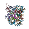
| Title | Structural mechanism of HP1⍺-dependent transcriptional repression and chromatin compaction. |
|---|---|
| Journal, issue, pages | Structure, Vol. 32, Issue 11, Page 2094-22106.e6, Year 2024 |
| Publish date | Nov 7, 2024 |
 Authors Authors | Vladyslava Sokolova / Jacob Miratsky / Vladimir Svetlov / Michael Brenowitz / John Vant / Tyler S Lewis / Kelly Dryden / Gahyun Lee / Shayan Sarkar / Evgeny Nudler / Abhishek Singharoy / Dongyan Tan /  |
| PubMed Abstract | Heterochromatin protein 1 (HP1) plays a central role in establishing and maintaining constitutive heterochromatin. However, the mechanisms underlying HP1-nucleosome interactions and their ...Heterochromatin protein 1 (HP1) plays a central role in establishing and maintaining constitutive heterochromatin. However, the mechanisms underlying HP1-nucleosome interactions and their contributions to heterochromatin functions remain elusive. Here, we present the cryoelectron microscopy (cryo-EM) structure of an HP1α dimer bound to an H2A.Z-nucleosome, revealing two distinct HP1α-nucleosome interfaces. The primary HP1α binding site is located at the N terminus of histone H3, specifically at the trimethylated lysine 9 (K9me3) region, while a secondary binding site is situated near histone H2B, close to nucleosome superhelical location 4 (SHL4). Our biochemical data further demonstrates that HP1α binding influences the dynamics of DNA on the nucleosome. It promotes DNA unwrapping near the nucleosome entry and exit sites while concurrently restricting DNA accessibility in the vicinity of SHL4. Our study offers a model for HP1α-mediated heterochromatin maintenance and gene silencing. It also sheds light on the H3K9me-independent role of HP1 in responding to DNA damage. |
 External links External links | Structure / PubMed:39383876 / PubMed Central |
| Methods | EM (single particle) |
| Resolution | 3.5 - 6.6 Å |
| Structure data |  EMDB-42690: Heterochromatin protein 1 (HP1) alpha in complex with an H2A.Z nucleosome(focused refined map)  EMDB-42692: K9me3-H2A.Z nucleosome from focus refinement of the HP1-H2A.Z nucleosome complex  EMDB-42773: structure of the K9me3-H2A.Z nucleosome EMDB-42774, PDB-8uxq: |
| Source |
|
 Keywords Keywords | STRUCTURAL PROTEIN / heterochromatin / nucleosome / chromatin / GENE REGULATION |


 homo sapiens (human)