 Movie
Movie Controller
 Structure viewers
 About Yorodumi Papers


+Search query
-Structure paper
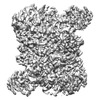
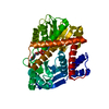
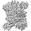
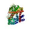
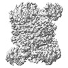
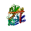
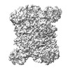
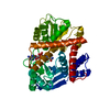
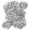
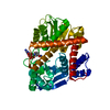
| Title | Coenzyme-binding pathway on glutamate dehydrogenase suggested from multiple-binding sites visualized by cryo-electron microscopy. |
|---|---|
| Journal, issue, pages | FEBS J, Vol. 290, Issue 23, Page 5514-5535, Year 2023 |
| Publish date | Sep 11, 2023 |
 Authors Authors | Taiki Wakabayashi / Mao Oide / Takayuki Kato / Masayoshi Nakasako /  |
| PubMed Abstract | The structure of hexameric glutamate dehydrogenase (GDH) in the presence of the coenzyme nicotinamide adenine dinucleotide phosphate (NADP) was visualized using cryogenic transmission electron ...The structure of hexameric glutamate dehydrogenase (GDH) in the presence of the coenzyme nicotinamide adenine dinucleotide phosphate (NADP) was visualized using cryogenic transmission electron microscopy to investigate the ligand-binding pathways to the active site of the enzyme. Each subunit of GDH comprises one hexamer-forming core domain and one nucleotide-binding domain (NAD domain), which spontaneously opens and closes the active-site cleft situated between the two domains. In the presence of NADP, the potential map of GDH hexamer, assuming D3 symmetry, was determined at a resolution of 2.4 Å, but the NAD domain was blurred due to the conformational variety. After focused classification with respect to the NAD domain, the potential maps interpreted as NADP molecules appeared at five different sites in the active-site cleft. The subunits associated with NADP molecules were close to one of the four metastable conformations in the unliganded state. Three of the five binding sites suggested a pathway of NADP molecules to approach the active-site cleft for initiating the enzymatic reaction. The other two binding modes may rarely appear in the presence of glutamate, as demonstrated by the reaction kinetics. Based on the visualized structures and the results from the enzymatic kinetics, we discussed the binding modes of NADP to GDH in the absence and presence of glutamate. |
 External links External links | FEBS J / PubMed:37682540 |
| Methods | EM (single particle) |
| Resolution | 3.08 - 3.29 Å |
| Structure data | EMDB-34805, PDB-8hho: EMDB-34826, PDB-8hiq: EMDB-34830, PDB-8hiz: EMDB-34831, PDB-8hj3: EMDB-34835, PDB-8hj9: |
| Chemicals |  ChemComp-NAP: |
| Source |
|
 Keywords Keywords | OXIDOREDUCTASE / Complex / Coenzyme / NADP |

 thermococcus profundus (archaea)