Ministry of Education, Culture, Sports, Science and Technology (Japan)
jp15076210
Japan
Ministry of Education, Culture, Sports, Science and Technology (Japan)
jp20050030
Japan
Ministry of Education, Culture, Sports, Science and Technology (Japan)
jp22018027
Japan
Ministry of Education, Culture, Sports, Science and Technology (Japan)
jp23120525
Japan
Ministry of Education, Culture, Sports, Science and Technology (Japan)
jp25120725
Japan
Ministry of Education, Culture, Sports, Science and Technology (Japan)
jp15H01647
Japan
Ministry of Education, Culture, Sports, Science and Technology (Japan)
jp17H05891
Japan
Japan Science and Technology
JPMJPR22E2
Japan
Citation
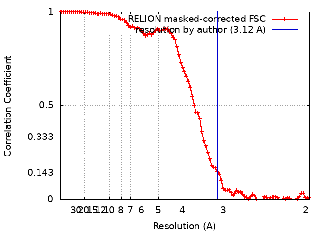
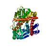
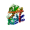
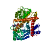
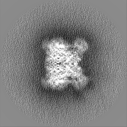
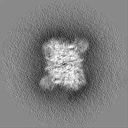
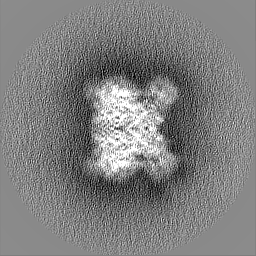
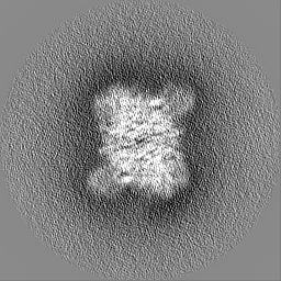
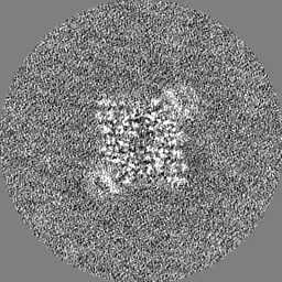
Journal: FEBS J / Year: 2023 Title: Coenzyme-binding pathway on glutamate dehydrogenase suggested from multiple-binding sites visualized by cryo-electron microscopy. Authors: Taiki Wakabayashi / Mao Oide / Takayuki Kato / Masayoshi Nakasako / Abstract: The structure of hexameric glutamate dehydrogenase (GDH) in the presence of the coenzyme nicotinamide adenine dinucleotide phosphate (NADP) was visualized using cryogenic transmission electron ...The structure of hexameric glutamate dehydrogenase (GDH) in the presence of the coenzyme nicotinamide adenine dinucleotide phosphate (NADP) was visualized using cryogenic transmission electron microscopy to investigate the ligand-binding pathways to the active site of the enzyme. Each subunit of GDH comprises one hexamer-forming core domain and one nucleotide-binding domain (NAD domain), which spontaneously opens and closes the active-site cleft situated between the two domains. In the presence of NADP, the potential map of GDH hexamer, assuming D3 symmetry, was determined at a resolution of 2.4 Å, but the NAD domain was blurred due to the conformational variety. After focused classification with respect to the NAD domain, the potential maps interpreted as NADP molecules appeared at five different sites in the active-site cleft. The subunits associated with NADP molecules were close to one of the four metastable conformations in the unliganded state. Three of the five binding sites suggested a pathway of NADP molecules to approach the active-site cleft for initiating the enzymatic reaction. The other two binding modes may rarely appear in the presence of glutamate, as demonstrated by the reaction kinetics. Based on the visualized structures and the results from the enzymatic kinetics, we discussed the binding modes of NADP to GDH in the absence and presence of glutamate.
In the structure databanks used in Yorodumi, some data are registered as the other names, "COVID-19 virus" and "2019-nCoV". Here are the details of the virus and the list of structure data.
Jan 31, 2019. EMDB accession codes are about to change! (news from PDBe EMDB page)
EMDB accession codes are about to change! (news from PDBe EMDB page)
The allocation of 4 digits for EMDB accession codes will soon come to an end. Whilst these codes will remain in use, new EMDB accession codes will include an additional digit and will expand incrementally as the available range of codes is exhausted. The current 4-digit format prefixed with “EMD-” (i.e. EMD-XXXX) will advance to a 5-digit format (i.e. EMD-XXXXX), and so on. It is currently estimated that the 4-digit codes will be depleted around Spring 2019, at which point the 5-digit format will come into force.
The EM Navigator/Yorodumi systems omit the EMD- prefix.
Related info.:Q: What is EMD? / ID/Accession-code notation in Yorodumi/EM Navigator
Yorodumi is a browser for structure data from EMDB, PDB, SASBDB, etc.
This page is also the successor to EM Navigator detail page, and also detail information page/front-end page for Omokage search.
The word "yorodu" (or yorozu) is an old Japanese word meaning "ten thousand". "mi" (miru) is to see.
Related info.:EMDB / PDB / SASBDB / Comparison of 3 databanks / Yorodumi Search / Aug 31, 2016. New EM Navigator & Yorodumi / Yorodumi Papers / Jmol/JSmol / Function and homology information / Changes in new EM Navigator and Yorodumi
 Movie
Movie Controller

 Yorodumi
 Open data
 Basic information
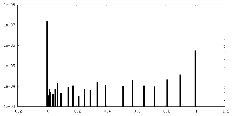
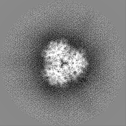
 Map data
 Sample
 Keywords
 Function and homology information
 Thermococcus profundus (archaea)
 Authors
 Japan, 14 items
  Citation
 Structure visualization
 Downloads & links
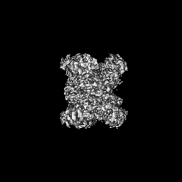
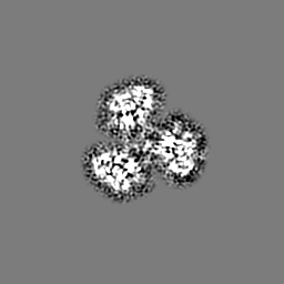
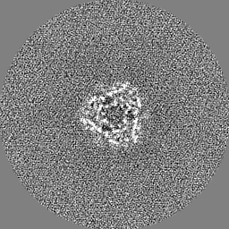
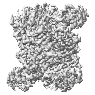 emd_34835.png
 http://ftp.pdbj.org/pub/emdb/structures/EMD-34835


 Z (Sec.)
 Y (Row.)
 X (Col.)


 Sample components

 Processing
 Electron microscopy
 FIELD EMISSION GUN