Ministry of Education, Culture, Sports, Science and Technology (Japan)
jp15076210
日本
Ministry of Education, Culture, Sports, Science and Technology (Japan)
jp20050030
日本
Ministry of Education, Culture, Sports, Science and Technology (Japan)
jp22018027
日本
Ministry of Education, Culture, Sports, Science and Technology (Japan)
jp23120525
日本
Ministry of Education, Culture, Sports, Science and Technology (Japan)
jp25120725
日本
Ministry of Education, Culture, Sports, Science and Technology (Japan)
jp15H01647
日本
Ministry of Education, Culture, Sports, Science and Technology (Japan)
jp17H05891
日本
Japan Science and Technology
JPMJPR22E2
日本
引用
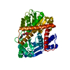
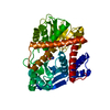
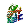
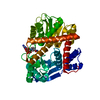
ジャーナル: FEBS J / 年: 2023 タイトル: Coenzyme-binding pathway on glutamate dehydrogenase suggested from multiple-binding sites visualized by cryo-electron microscopy. 著者: Taiki Wakabayashi / Mao Oide / Takayuki Kato / Masayoshi Nakasako / 要旨: The structure of hexameric glutamate dehydrogenase (GDH) in the presence of the coenzyme nicotinamide adenine dinucleotide phosphate (NADP) was visualized using cryogenic transmission electron ...The structure of hexameric glutamate dehydrogenase (GDH) in the presence of the coenzyme nicotinamide adenine dinucleotide phosphate (NADP) was visualized using cryogenic transmission electron microscopy to investigate the ligand-binding pathways to the active site of the enzyme. Each subunit of GDH comprises one hexamer-forming core domain and one nucleotide-binding domain (NAD domain), which spontaneously opens and closes the active-site cleft situated between the two domains. In the presence of NADP, the potential map of GDH hexamer, assuming D3 symmetry, was determined at a resolution of 2.4 Å, but the NAD domain was blurred due to the conformational variety. After focused classification with respect to the NAD domain, the potential maps interpreted as NADP molecules appeared at five different sites in the active-site cleft. The subunits associated with NADP molecules were close to one of the four metastable conformations in the unliganded state. Three of the five binding sites suggested a pathway of NADP molecules to approach the active-site cleft for initiating the enzymatic reaction. The other two binding modes may rarely appear in the presence of glutamate, as demonstrated by the reaction kinetics. Based on the visualized structures and the results from the enzymatic kinetics, we discussed the binding modes of NADP to GDH in the absence and presence of glutamate.
履歴
登録
2022年11月21日
登録サイト: PDBJ / 処理サイト: PDBJ
改定 1.0
2023年2月8日
Provider: repository / タイプ: Initial release
改定 2.0
2023年8月9日
Group: Atomic model / Derived calculations / Structure summary カテゴリ: atom_site / pdbx_contact_author / struct_conf Item: _atom_site.B_iso_or_equiv / _atom_site.Cartn_x ..._atom_site.B_iso_or_equiv / _atom_site.Cartn_x / _atom_site.Cartn_y / _atom_site.Cartn_z / _atom_site.pdbx_formal_charge / _struct_conf.beg_auth_comp_id / _struct_conf.beg_auth_seq_id / _struct_conf.beg_label_comp_id / _struct_conf.beg_label_seq_id / _struct_conf.pdbx_PDB_helix_length 解説: Atomic clashes 詳細: All close contacts between heavy atoms were removed. Provider: author / タイプ: Coordinate replacement
 ムービー
ムービー コントローラー


 データを開く
 基本情報
基本情報 要素
要素 キーワード
キーワード 機能・相同性情報
 Thermococcus profundus (古細菌)
 データ登録者
 日本, 14件
  引用
引用 構造の表示
構造の表示 ダウンロードとリンク
 その他のダウンロード


 PDBj
PDBj 集合体
集合体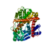


 分子量: 743.405 Da / 分子数: 1 / 由来タイプ: 合成 / 式: C21H28N7O17P3 / タイプ: SUBJECT OF INVESTIGATION
 試料調製
試料調製 電子顕微鏡撮影
 FIELD EMISSION GUN / 加速電圧: 300 kV / 照射モード: OTHER
 解析
解析