 Movie
Movie Controller

[English] 日本語
 Yorodumi
- PDB-8hj3: cryoEM structure of glutamate dehydrogenase from Thermococcus pro... -
+ Open data

- Basic information
| Entry | Database: PDB / ID: 8hj3 | |||||||||||||||||||||||||||||||||||||||||||||
|---|---|---|---|---|---|---|---|---|---|---|---|---|---|---|---|---|---|---|---|---|---|---|---|---|---|---|---|---|---|---|---|---|---|---|---|---|---|---|---|---|---|---|---|---|---|---|
| Title | cryoEM structure of glutamate dehydrogenase from Thermococcus profundus in complex with NADP | |||||||||||||||||||||||||||||||||||||||||||||
 Components Components | Glutamate dehydrogenase | |||||||||||||||||||||||||||||||||||||||||||||
 Keywords Keywords | OXIDOREDUCTASE / Complex / Coenzyme / NADP | |||||||||||||||||||||||||||||||||||||||||||||
| Function / homology |  Function and homology information Function and homology informationglutamate dehydrogenase [NAD(P)+] / L-glutamate dehydrogenase (NADP+) activity / L-glutamate dehydrogenase (NAD+) activity / L-glutamate catabolic process Similarity search - Function | |||||||||||||||||||||||||||||||||||||||||||||
| Biological species |   Thermococcus profundus (archaea) Thermococcus profundus (archaea) | |||||||||||||||||||||||||||||||||||||||||||||
| Method | ELECTRON MICROSCOPY / single particle reconstruction / cryo EM / Resolution: 3.29 Å | |||||||||||||||||||||||||||||||||||||||||||||
 Authors Authors | Wakabayashi, T. / Oide, M. / Kato, T. / Nakasako, M. | |||||||||||||||||||||||||||||||||||||||||||||
| Funding support |  Japan, 14items Japan, 14items
| |||||||||||||||||||||||||||||||||||||||||||||
 Citation Citation | Journal: FEBS J / Year: 2023 Title: Coenzyme-binding pathway on glutamate dehydrogenase suggested from multiple-binding sites visualized by cryo-electron microscopy. Authors: Taiki Wakabayashi / Mao Oide / Takayuki Kato / Masayoshi Nakasako / Abstract: The structure of hexameric glutamate dehydrogenase (GDH) in the presence of the coenzyme nicotinamide adenine dinucleotide phosphate (NADP) was visualized using cryogenic transmission electron ...The structure of hexameric glutamate dehydrogenase (GDH) in the presence of the coenzyme nicotinamide adenine dinucleotide phosphate (NADP) was visualized using cryogenic transmission electron microscopy to investigate the ligand-binding pathways to the active site of the enzyme. Each subunit of GDH comprises one hexamer-forming core domain and one nucleotide-binding domain (NAD domain), which spontaneously opens and closes the active-site cleft situated between the two domains. In the presence of NADP, the potential map of GDH hexamer, assuming D3 symmetry, was determined at a resolution of 2.4 Å, but the NAD domain was blurred due to the conformational variety. After focused classification with respect to the NAD domain, the potential maps interpreted as NADP molecules appeared at five different sites in the active-site cleft. The subunits associated with NADP molecules were close to one of the four metastable conformations in the unliganded state. Three of the five binding sites suggested a pathway of NADP molecules to approach the active-site cleft for initiating the enzymatic reaction. The other two binding modes may rarely appear in the presence of glutamate, as demonstrated by the reaction kinetics. Based on the visualized structures and the results from the enzymatic kinetics, we discussed the binding modes of NADP to GDH in the absence and presence of glutamate. | |||||||||||||||||||||||||||||||||||||||||||||
| History |
|
- Structure visualization
| Structure viewer | Molecule: MolmilJmol/JSmol |
|---|
- Downloads & links
-Download
| PDBx/mmCIF format | 8hj3.cif.gz | 85.6 KB | Display | PDBx/mmCIF format |
|---|---|---|---|---|
| PDB format | pdb8hj3.ent.gz | 62.4 KB | Display | PDB format |
| PDBx/mmJSON format | 8hj3.json.gz | Tree view | PDBx/mmJSON format | |
| Others |  Other downloads Other downloads |
-Validation report
| Arichive directory | https://data.pdbj.org/pub/pdb/validation_reports/hj/8hj3ftp://data.pdbj.org/pub/pdb/validation_reports/hj/8hj3 | HTTPS FTP |
|---|
-Related structure data
| Related structure data |  34831MC 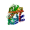 8hhoC 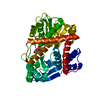 8hiqC 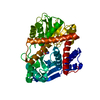 8hizC 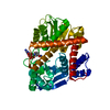 8hj9C C: citing same article ( M: map data used to model this data |
|---|---|
| Similar structure data |


-Links
 PDBj
PDBj- Assembly
| Deposited unit | 
|
|---|---|
| 1 |
|
-Components
| #1: Protein | Mass: 46758.477 Da / Num. of mol.: 1 Source method: isolated from a genetically manipulated source Source: (gene. exp.) Thermococcus profundus (archaea) / Gene: gdhA / Production host:  References: UniProt: O74024, glutamate dehydrogenase [NAD(P)+] |
|---|---|
| #2: Chemical | ChemComp-NAP /   Mass: 743.405 Da / Num. of mol.: 1 / Source method: obtained synthetically / Formula: C21H28N7O17P3 / Feature type: SUBJECT OF INVESTIGATION Mass: 743.405 Da / Num. of mol.: 1 / Source method: obtained synthetically / Formula: C21H28N7O17P3 / Feature type: SUBJECT OF INVESTIGATION |
| Has ligand of interest | Y |
-Experimental details
-Experiment
| Experiment | Method: ELECTRON MICROSCOPY |
|---|---|
| EM experiment | Aggregation state: PARTICLE / 3D reconstruction method: single particle reconstruction |
- Sample preparation
| Component | Name: Hexamer of glutamate dehydrogenase in the presence of NADP Type: COMPLEX / Entity ID: #1 / Source: RECOMBINANT | ||||||||||||||||||||
|---|---|---|---|---|---|---|---|---|---|---|---|---|---|---|---|---|---|---|---|---|---|
| Molecular weight | Value: 0.264 MDa / Experimental value: YES | ||||||||||||||||||||
| Source (natural) | Organism: Thermococcus profundus (archaea) | ||||||||||||||||||||
| Source (recombinant) | Organism: | ||||||||||||||||||||
| Buffer solution | pH: 7.5 | ||||||||||||||||||||
| Buffer component |
| ||||||||||||||||||||
| Specimen | Conc.: 0.0036 mg/ml / Embedding applied: NO / Shadowing applied: NO / Staining applied: NO / Vitrification applied: YES | ||||||||||||||||||||
| Specimen support | Grid material: MOLYBDENUM / Grid mesh size: 200 divisions/in. / Grid type: Quantifoil R1.2/1.3 | ||||||||||||||||||||
| Vitrification | Instrument: FEI VITROBOT MARK IV / Cryogen name: ETHANE / Humidity: 100 % / Chamber temperature: 277 K |
- Electron microscopy imaging
| Microscopy | Model: JEOL CRYO ARM 300 |
|---|---|
| Electron gun | Electron source:  FIELD EMISSION GUN / Accelerating voltage: 300 kV / Illumination mode: OTHER FIELD EMISSION GUN / Accelerating voltage: 300 kV / Illumination mode: OTHER |
| Electron lens | Mode: BRIGHT FIELD / Nominal defocus max: 3300 nm / Nominal defocus min: 500 nm |
| Specimen holder | Cryogen: NITROGEN |
| Image recording | Electron dose: 1.2 e/Å2 / Film or detector model: GATAN K3 (6k x 4k) / Num. of real images: 10389 |
- Processing
| EM software |
| ||||||||||||||||||||||||||||||||
|---|---|---|---|---|---|---|---|---|---|---|---|---|---|---|---|---|---|---|---|---|---|---|---|---|---|---|---|---|---|---|---|---|---|
| CTF correction | Type: PHASE FLIPPING AND AMPLITUDE CORRECTION | ||||||||||||||||||||||||||||||||
| Particle selection | Num. of particles selected: 3037558 | ||||||||||||||||||||||||||||||||
| Symmetry | Point symmetry: C1 (asymmetric) | ||||||||||||||||||||||||||||||||
| 3D reconstruction | Resolution: 3.29 Å / Resolution method: FSC 0.143 CUT-OFF / Num. of particles: 59423 / Symmetry type: POINT | ||||||||||||||||||||||||||||||||
| Atomic model building | Protocol: RIGID BODY FIT |
