 Movie
Movie Controller
 Structure viewers
 About Yorodumi Papers


+Search query
-Structure paper
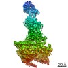
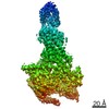
| Title | Cryo-EM structures of PAC1 receptor reveal ligand binding mechanism. |
|---|---|
| Journal, issue, pages | Cell Res, Vol. 30, Issue 5, Page 436-445, Year 2020 |
| Publish date | Feb 11, 2020 |
 Authors Authors | Jia Wang / Xianqiang Song / Dandan Zhang / Xiaoqing Chen / Xun Li / Yaping Sun / Cui Li / Yunpeng Song / Yao Ding / Ruobing Ren / Essa Hu Harrington / Liaoyuan A Hu / Wenge Zhong / Cen Xu / Xin Huang / Hong-Wei Wang / Yingli Ma /   |
| PubMed Abstract | The pituitary adenylate cyclase-activating polypeptide type I receptor (PAC1R) belongs to the secretin receptor family and is widely distributed in the central neural system and peripheral organs. ...The pituitary adenylate cyclase-activating polypeptide type I receptor (PAC1R) belongs to the secretin receptor family and is widely distributed in the central neural system and peripheral organs. Abnormal activation of the receptor mediates trigeminovascular activation and sensitization, which is highly related to migraine, making PAC1R a potential therapeutic target. Elucidation of PAC1R activation mechanism would benefit discovery of therapeutic drugs for neuronal disorders. PAC1R activity is governed by pituitary adenylate cyclase-activating polypeptide (PACAP), known as a major vasodilator neuropeptide, and maxadilan, a native peptide from the sand fly, which is also capable of activating the receptor with similar potency. These peptide ligands have divergent sequences yet initiate convergent PAC1R activity. It is of interest to understand the mechanism of PAC1R ligand recognition and receptor activity regulation through structural biology. Here we report two near-atomic resolution cryo-EM structures of PAC1R activated by PACAP38 or maxadilan, providing structural insights into two distinct ligand binding modes. The structures illustrate flexibility of the extracellular domain (ECD) for ligands with distinct conformations, where ECD accommodates ligands in different orientations while extracellular loop 1 (ECL1) protrudes to further anchor the ligand bound in the orthosteric site. By structure-guided molecular modeling and mutagenesis, we tested residues in the ligand-binding pockets and identified clusters of residues that are critical for receptor activity. The structures reported here for the first time elucidate the mechanism of specificity and flexibility of ligand recognition and binding for PAC1R, and provide insights toward the design of therapeutic molecules targeting PAC1R. |
 External links External links | Cell Res / PubMed:32047270 / PubMed Central |
| Methods | EM (single particle) |
| Resolution | 3.5 - 3.6 Å |
| Structure data | EMDB-30047, PDB-6m1h: EMDB-30048, PDB-6m1i: |
| Source |
|
 Keywords Keywords | PROTEIN BINDING / GPCR |


 homo sapiens (human)
 lutzomyia longipalpis (insect)
