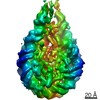
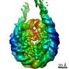
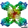
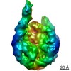
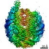
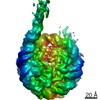
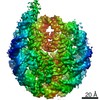
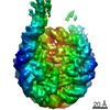
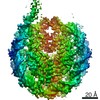
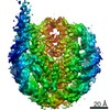
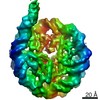
Structural heterogeneity of nucleosomes in functional chromosomes is unknown. Here, we devise the template-, reference- and selection-free (TRSF) cryo-EM pipeline to simultaneously reconstruct cryo- ...Structural heterogeneity of nucleosomes in functional chromosomes is unknown. Here, we devise the template-, reference- and selection-free (TRSF) cryo-EM pipeline to simultaneously reconstruct cryo-EM structures of protein complexes from interphase or metaphase chromosomes. The reconstructed interphase and metaphase nucleosome structures are on average indistinguishable from canonical nucleosome structures, despite DNA sequence heterogeneity, cell-cycle-specific posttranslational modifications, and interacting proteins. Nucleosome structures determined by a decoy-classifying method and structure variability analyses reveal the nucleosome structural variations in linker DNA, histone tails, and nucleosome core particle configurations, suggesting that the opening of linker DNA, which is correlated with H2A C-terminal tail positioning, is suppressed in chromosomes. High-resolution (3.4-3.5 Å) nucleosome structures indicate DNA-sequence-independent stabilization of superhelical locations ±0-1 and ±3.5-4.5. The linker histone H1.8 preferentially binds to metaphase chromatin, from which chromatosome cryo-EM structures with H1.8 at the on-dyad position are reconstituted. This study presents the structural characteristics of nucleosomes in chromosomes.
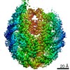
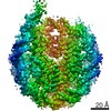
EMDB-22790, PDB-7kbd: Nucleosome in interphase chromosome formed in Xenopus egg extract (oligo fraction) Method: EM (single particle) / Resolution: 3.38 Å
EMDB-22791, PDB-7kbe: Nucleosome isolated from metaphase chromosome formed in Xenopus egg extract (oligo fraction) Method: EM (single particle) / Resolution: 3.5 Å
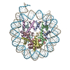
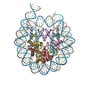
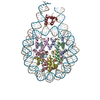
EMDB-22792, PDB-7kbf: H1.8 bound nucleosome isolated from metaphase chromosome in Xenopus egg extract (oligo fraction) Method: EM (single particle) / Resolution: 4.42 Å
EMDB-22793: Nucleosome isolated from interphase chromosome formed in Xenopus egg extract (oligo fraction) (unbiased reconstruction) Method: EM (single particle) / Resolution: 4.2 Å
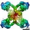
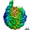
EMDB-22794: Alpha2-macroglobulin family protein isolated from interphase chromosome formed in Xenopus egg extract Method: EM (single particle) / Resolution: 8.07 Å
EMDB-22795: Nucleosome isolated from metaphase chromosome formed in Xenopus egg extract (oligo fraction) (unbiased reconstruction) Method: EM (single particle) / Resolution: 4.2 Å
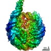
EMDB-22796: Alpha2-macroglobulin family protein isolated from metaphase chromosome formed in Xenopus egg extract Method: EM (single particle) / Resolution: 5.5 Å
EMDB-22797: Nucleosome isolated from interphase chromosome formed in Xenopus egg extract (mono fraction) Method: EM (single particle) / Resolution: 5.12 Å
EMDB-22798: Nucleosome isolated from interphase chromosome formed in Xenopus egg extract (di fraction) Method: EM (single particle) / Resolution: 4.74 Å
EMDB-22800: Nucleosome isolated from metaphase chromosome formed in Xenopus egg extract (mono fraction) Method: EM (single particle) / Resolution: 5.64 Å
EMDB-22801: Nucleosome isolated from metaphase chromosome formed in Xenopus egg extract (di fraction) Method: EM (single particle) / Resolution: 8.1 Å
EMDB-23819: Nucleosome isolated from the interphase chromosome (oligo fraction, egg extract lot 2) Method: EM (single particle) / Resolution: 3.54 Å
EMDB-23820: Nucleosome isolated from the interphase chromosome (oligo fraction, egg extract lot 2, simultaneous structures reconstruction) Method: EM (single particle) / Resolution: 4.42 Å
EMDB-23821: Nucleosome isolated from the metaphase chromosome (oligo fraction, egg extract lot 2) Method: EM (single particle) / Resolution: 3.77 Å
EMDB-23822: Nucleosome isolated from the metaphase chromosome (oligo fraction, egg extract lot 2, simultaneous structures reconstruction) Method: EM (single particle) / Resolution: 4.32 Å
EMDB-23823: Closed linker DNA nucleosome reconstituted with GUB DNA Method: EM (single particle) / Resolution: 3.77 Å
EMDB-23824: Open linker DNA nucleosome reconstituted with GUB DNA Method: EM (single particle) / Resolution: 4.52 Å
EMDB-23826: Nucleosome isolated from interphase chromosome formed in Xenopus egg extract (crosslinked after MNase, egg extract lot 2) Method: EM (single particle) / Resolution: 5.44 Å
Source
xenopus laevis (African clawed frog)
synthetic construct (others)
Keywords
STRUCTURAL PROTEIN/DNA / nucleosome / interphase / cell cycle / chromatin / DNA BINDING PROTEIN / STRUCTURAL PROTEIN-DNA complex / DNA BINDING PROTEIN/DNA / M phase / Xenopus egg extract / DNA BINDING PROTEIN-DNA complex / H1 / Chromatosome / Linker histone
+
About Yorodumi Papers
-
News
-
Feb 9, 2022. New format data for meta-information of EMDB entries
New format data for meta-information of EMDB entries
Version 3 of the EMDB header file is now the official format.
The previous official version 1.9 will be removed from the archive.
In the structure databanks used in Yorodumi, some data are registered as the other names, "COVID-19 virus" and "2019-nCoV". Here are the details of the virus and the list of structure data.
Jan 31, 2019. EMDB accession codes are about to change! (news from PDBe EMDB page)
EMDB accession codes are about to change! (news from PDBe EMDB page)
The allocation of 4 digits for EMDB accession codes will soon come to an end. Whilst these codes will remain in use, new EMDB accession codes will include an additional digit and will expand incrementally as the available range of codes is exhausted. The current 4-digit format prefixed with “EMD-” (i.e. EMD-XXXX) will advance to a 5-digit format (i.e. EMD-XXXXX), and so on. It is currently estimated that the 4-digit codes will be depleted around Spring 2019, at which point the 5-digit format will come into force.
The EM Navigator/Yorodumi systems omit the EMD- prefix.
Related info.:Q: What is EMD? / ID/Accession-code notation in Yorodumi/EM Navigator
 Movie
Movie Controller
 Structure viewers
 About Yorodumi Papers


 Authors
 External links
 Keywords