 Movie
Movie Controller
 Structure viewers
 About Yorodumi Papers


+Search query
-Structure paper
| Title | Structural insights into context-dependent inhibitory mechanisms of chloramphenicol in cells. |
|---|---|
| Journal, issue, pages | Nat Struct Mol Biol, Vol. 32, Issue 2, Page 257-267, Year 2025 |
| Publish date | Dec 12, 2024 |
 Authors Authors | Liang Xue / Christian M T Spahn / Magdalena Schacherl / Julia Mahamid /   |
| PubMed Abstract | Ribosome-targeting antibiotics represent an important class of antimicrobial drugs. Chloramphenicol (Cm) is a well-studied ribosomal peptidyl transferase center (PTC) binder and growing evidence ...Ribosome-targeting antibiotics represent an important class of antimicrobial drugs. Chloramphenicol (Cm) is a well-studied ribosomal peptidyl transferase center (PTC) binder and growing evidence suggests that its inhibitory action depends on the sequence of the nascent peptide. How such selective inhibition on the molecular scale manifests on the cellular level remains unclear. Here, we use cryo-electron tomography to analyze the impact of Cm inside the bacterium Mycoplasma pneumoniae. By resolving the Cm-bound ribosomes to 3.0 Å, we elucidate Cm's coordination with natural nascent peptides and transfer RNAs in the PTC. We find that Cm leads to the accumulation of a number of translation elongation states, indicating ongoing futile accommodation cycles, and to extensive ribosome collisions. We, thus, suggest that, beyond its direct inhibition of protein synthesis, the action of Cm may involve the activation of cellular stress responses. This work exemplifies how in-cell structural biology can expand the understanding of mechanisms of action for extensively studied antibiotics. |
 External links External links | Nat Struct Mol Biol / PubMed:39668257 / PubMed Central |
| Methods | EM (subtomogram averaging) |
| Resolution | 2.9 - 15.0 Å |
| Structure data | EMDB-17132: 3.0Angstrom 70S ribosome map in chloramphenicol-treated Mycoplasma pneumoniae cells EMDB-17133: 3.2Angstrom 30S ribosome focused-refined map in chloramphenicol-treated Mycoplasma pneumoniae cells EMDB-17134: 2.9Angstrom 50S ribosome focused-refined map in chloramphenicol-treated Mycoplasma pneumoniae cells EMDB-17135: 70S ribosome with additional S4-LSU in chloramphenicol-treated Mycoplasma pneumoniae cells 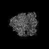 EMDB-17136: 70S ribosome with A and P-site tRNAs in chloramphenicol-treated Mycoplasma pneumoniae cells, K3 data 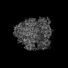 EMDB-17137: 70S ribosome with A, P and E-site tRNAs in chloramphenicol-treated Mycoplasma pneumoniae cells, K3 data 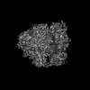 EMDB-17138: 70S ribosome with a and P-site tRNAs in chloramphenicol-treated Mycoplasma pneumoniae cells, K3 data 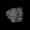 EMDB-17139: 70S ribosome with a, P and E-site tRNAs in chloramphenicol-treated Mycoplasma pneumoniae cells, K3 data  EMDB-17140: 70S ribosome with EF-Tu-tRNA and P-site tRNA in chloramphenicol-treated Mycoplasma pneumoniae cells, K3 data 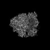 EMDB-17141: 70S ribosome with EF-Tu-tRNA, P and E-site tRNAs in chloramphenicol-treated Mycoplasma pneumoniae cells, K3 data 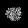 EMDB-17142: 70S ribosome with A* and P/E-site tRNAs in chloramphenicol-treated Mycoplasma pneumoniae cells, K3 data  EMDB-17143: polysome/di-ribosome class I in chloramphenicol-treated Mycoplasma pneumoniae cells  EMDB-17144: polysome/di-ribosome class II in chloramphenicol-treated Mycoplasma pneumoniae cells EMDB-17145: polysome/di-ribosome class III in chloramphenicol-treated Mycoplasma pneumoniae cells  EMDB-17146: 70S ribosome with additional S4-LSU in native untreated Mycoplasma pneumoniae cells  EMDB-17147: free 50S with additional S4-LSU in native untreated Mycoplasma pneumoniae cells |
| Chemicals |  ChemComp-SPD:  ChemComp-PUT:  ChemComp-N2P:  ChemComp-MG:  ChemComp-ZN:  ChemComp-HOH: 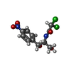 ChemComp-CLM:  ChemComp-K:  ChemComp-SPM:  ChemComp-LYS: |
| Source |
|
 Keywords Keywords | TRANSLATION / In situ / Ribosome / Chloramphenicol / cryo-ET |


 mycoplasmoides pneumoniae m129 (bacteria)