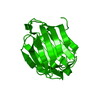
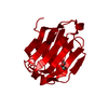
| タイトル | Galectin-10: a new structural type of prototype galectin dimer and effects on saccharide ligand binding. |
|---|
| ジャーナル・号・ページ | Glycobiology, Vol. 28, Page 159-168, Year 2018 |
|---|
| 掲載日 | 2017年6月8日 (構造データの登録日) |
|---|
 著者 著者 | Su, J. / Gao, J. / Si, Y. / Cui, L. / Song, C. / Wang, Y. / Wu, R. / Tai, G. / Zhou, Y. |
|---|
 リンク リンク |  Glycobiology / Glycobiology /  PubMed:29293962 PubMed:29293962 |
|---|
| 手法 | X線回折 |
|---|
| 解像度 | 1.55 - 2.004 Å |
|---|
| 構造データ | PDB-5xrg:
Galectin-10/Charcot-Leyden crystal protein crystal structure
手法: X-RAY DIFFRACTION / 解像度: 1.55 Å PDB-5xrh:
Galectin-10/Charcot-Leyden crystal protein crystal structure
手法: X-RAY DIFFRACTION / 解像度: 1.55 Å PDB-5xri:
Galectin-10/Charcot-Leyden crystal protein-C29A crystal structure
手法: X-RAY DIFFRACTION / 解像度: 1.68 Å PDB-5xrj:
Galectin-10/Charcot-Leyden crystal protein variant H53A crystal structure
手法: X-RAY DIFFRACTION / 解像度: 1.9 Å PDB-5xrk:
Galectin-10/Charcot-Leyden crystal protein variant C57A crystal structure
手法: X-RAY DIFFRACTION / 解像度: 1.7 Å PDB-5xrl:
Galectin-10/Charcot-Leyden crystal protein variant N65A crystal structure
手法: X-RAY DIFFRACTION / 解像度: 2.004 Å PDB-5xrm:
Galectin-10/Charcot-Leyden crystal protein variant W72A crystal structure
手法: X-RAY DIFFRACTION / 解像度: 1.998 Å PDB-5xrn:
Galectin-10/Charcot-Leyden crystal protein variant K73A crystal structure
手法: X-RAY DIFFRACTION / 解像度: 1.6 Å PDB-5xro:
Galectin-10/Charcot-Leyden crystal protein variant Q74A crystal structure
手法: X-RAY DIFFRACTION / 解像度: 1.6 Å PDB-5xrp:
Galectin-10/Charcot-Leyden crystal protein variant Q75A
手法: X-RAY DIFFRACTION / 解像度: 2.001 Å PDB-5yt4:
Galectin-10 variant H53A soaked in glycerol for 5 minutes
手法: X-RAY DIFFRACTION / 解像度: 2 Å |
|---|
| 化合物 | |
|---|
| 由来 |  homo sapiens (ヒト) homo sapiens (ヒト)
|
|---|
 キーワード キーワード | PROTEIN BINDING / Galectin-10/Charcot-Leyden crystal protein / CELL ADHESION / Galectin-10 |
|---|
 著者
著者 リンク
リンク Glycobiology /
  PubMed:29293962


 キーワード
キーワード ムービー
ムービー コントローラー
 構造ビューア
 万見文献について


 homo sapiens (ヒト)