[English] 日本語
Yorodumi Papers
- Database of articles cited by EMDB/PDB/SASBDB data -

+
Search query

Keywords
Structure methods
Author
Journal
IF

-
Structure paper

TitleCoenzyme-binding pathway on glutamate dehydrogenase suggested from multiple-binding sites visualized by cryo-electron microscopy.
Journal, issue, pagesFEBS J, Vol. 290, Issue 23, Page 5514-5535, Year 2023
Publish dateSep 11, 2023
AuthorsTaiki Wakabayashi / Mao Oide / Takayuki Kato / Masayoshi Nakasako /
PubMed AbstractThe structure of hexameric glutamate dehydrogenase (GDH) in the presence of the coenzyme nicotinamide adenine dinucleotide phosphate (NADP) was visualized using cryogenic transmission electron ...The structure of hexameric glutamate dehydrogenase (GDH) in the presence of the coenzyme nicotinamide adenine dinucleotide phosphate (NADP) was visualized using cryogenic transmission electron microscopy to investigate the ligand-binding pathways to the active site of the enzyme. Each subunit of GDH comprises one hexamer-forming core domain and one nucleotide-binding domain (NAD domain), which spontaneously opens and closes the active-site cleft situated between the two domains. In the presence of NADP, the potential map of GDH hexamer, assuming D3 symmetry, was determined at a resolution of 2.4 Å, but the NAD domain was blurred due to the conformational variety. After focused classification with respect to the NAD domain, the potential maps interpreted as NADP molecules appeared at five different sites in the active-site cleft. The subunits associated with NADP molecules were close to one of the four metastable conformations in the unliganded state. Three of the five binding sites suggested a pathway of NADP molecules to approach the active-site cleft for initiating the enzymatic reaction. The other two binding modes may rarely appear in the presence of glutamate, as demonstrated by the reaction kinetics. Based on the visualized structures and the results from the enzymatic kinetics, we discussed the binding modes of NADP to GDH in the absence and presence of glutamate.
External linksFEBS J / PubMed:37682540
MethodsEM (single particle)
Resolution3.08 - 3.29 Å
Structure data

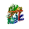
EMDB-34805, PDB-8hho:
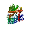
cryoEM structure of glutamate dehydrogenase from Thermococcus profundus in complex with NADP
Method: EM (single particle) / Resolution: 3.2 Å

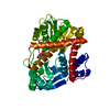
EMDB-34826, PDB-8hiq:
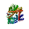
cryoEM structure of glutamate dehydrogenase from Thermococcus profundus in complex with NADP
Method: EM (single particle) / Resolution: 3.2 Å

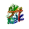
EMDB-34830, PDB-8hiz:
cryoEM structure of glutamate dehydrogenase from Thermococcus profundus in complex with NADP
Method: EM (single particle) / Resolution: 3.08 Å

EMDB-34831, PDB-8hj3:
cryoEM structure of glutamate dehydrogenase from Thermococcus profundus in complex with NADP
Method: EM (single particle) / Resolution: 3.29 Å

EMDB-34835, PDB-8hj9:
cryoEM structure of glutamate dehydrogenase from Thermococcus profundus in complex with NADP
Method: EM (single particle) / Resolution: 3.12 Å

Chemicals

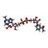
ChemComp-NAP:
NADP NICOTINAMIDE-ADENINE-DINUCLEOTIDE PHOSPHATE

Source
  • thermococcus profundus (archaea)
KeywordsOXIDOREDUCTASE / Complex / Coenzyme / NADP

+
About Yorodumi Papers

-
News

-
Feb 9, 2022. New format data for meta-information of EMDB entries

New format data for meta-information of EMDB entries

  • Version 3 of the EMDB header file is now the official format.
  • The previous official version 1.9 will be removed from the archive.

Related info.:EMDB header

External links:wwPDB to switch to version 3 of the EMDB data model

-
Aug 12, 2020. Covid-19 info

Covid-19 info

URL: https://pdbj.org/emnavi/covid19.php

New page: Covid-19 featured information page in EM Navigator.

Related info.:Covid-19 info / Mar 5, 2020. Novel coronavirus structure data

+
Mar 5, 2020. Novel coronavirus structure data

Novel coronavirus structure data

Related info.:Yorodumi Speices / Aug 12, 2020. Covid-19 info

External links:COVID-19 featured content - PDBj / Molecule of the Month (242):Coronavirus Proteases

+
Jan 31, 2019. EMDB accession codes are about to change! (news from PDBe EMDB page)

EMDB accession codes are about to change! (news from PDBe EMDB page)

  • The allocation of 4 digits for EMDB accession codes will soon come to an end. Whilst these codes will remain in use, new EMDB accession codes will include an additional digit and will expand incrementally as the available range of codes is exhausted. The current 4-digit format prefixed with “EMD-” (i.e. EMD-XXXX) will advance to a 5-digit format (i.e. EMD-XXXXX), and so on. It is currently estimated that the 4-digit codes will be depleted around Spring 2019, at which point the 5-digit format will come into force.
  • The EM Navigator/Yorodumi systems omit the EMD- prefix.

Related info.:Q: What is EMD? / ID/Accession-code notation in Yorodumi/EM Navigator

External links:EMDB Accession Codes are Changing Soon! / Contact to PDBj

+
Jul 12, 2017. Major update of PDB

Major update of PDB

  • wwPDB released updated PDB data conforming to the new PDBx/mmCIF dictionary.
  • This is a major update changing the version number from 4 to 5, and with Remediation, in which all the entries are updated.
  • In this update, many items about electron microscopy experimental information are reorganized (e.g. em_software).
  • Now, EM Navigator and Yorodumi are based on the updated data.

External links:wwPDB Remediation / Enriched Model Files Conforming to OneDep Data Standards Now Available in the PDB FTP Archive

-
Yorodumi Papers

Database of articles cited by EMDB/PDB/SASBDB data

  • Database of articles cited by EMDB, PDB, and SASBDB entries
  • Using PubMed data

Related info.:EMDB / PDB / SASBDB / Yorodumi / EMN Papers / Changes in new EM Navigator and Yorodumi

Read more