+Search query
-Structure paper
| Title | Structural basis of peptide recognition and activation of endothelin receptors. |
|---|---|
| Journal, issue, pages | Nat Commun, Vol. 14, Issue 1, Page 1268, Year 2023 |
| Publish date | Mar 7, 2023 |
 Authors Authors | Yujie Ji / Jia Duan / Qingning Yuan / Xinheng He / Gong Yang / Shengnan Zhu / Kai Wu / Wen Hu / Tianyu Gao / Xi Cheng / Hualiang Jiang / H Eric Xu / Yi Jiang /  |
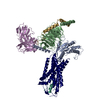
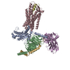
| PubMed Abstract | Endothelin system comprises three endogenous 21-amino-acid peptide ligands endothelin-1, -2, and -3 (ET-1/2/3), and two G protein-coupled receptor (GPCR) subtypes-endothelin receptor A (ETR) and B ...Endothelin system comprises three endogenous 21-amino-acid peptide ligands endothelin-1, -2, and -3 (ET-1/2/3), and two G protein-coupled receptor (GPCR) subtypes-endothelin receptor A (ETR) and B (ETR). Since ET-1, the first endothelin, was identified in 1988 as one of the most potent endothelial cell-derived vasoconstrictor peptides with long-lasting actions, the endothelin system has attracted extensive attention due to its critical role in vasoregulation and close relevance in cardiovascular-related diseases. Here we present three cryo-electron microscopy structures of ETR and ETR bound to ET-1 and ETR bound to the selective peptide IRL1620. These structures reveal a highly conserved recognition mode of ET-1 and characterize the ligand selectivity by ETRs. They also present several conformation features of the active ETRs, thus revealing a specific activation mechanism. Together, these findings deepen our understanding of endothelin system regulation and offer an opportunity to design selective drugs targeting specific ETR subtypes. |
 External links External links |  Nat Commun / Nat Commun /  PubMed:36882417 / PubMed:36882417 /  PubMed Central PubMed Central |
| Methods | EM (single particle) |
| Resolution | 2.99 - 3.5 Å |
| Structure data | EMDB-34619, PDB-8hbd: EMDB-34663, PDB-8hcq: EMDB-34667, PDB-8hcx: |
| Source |
|
 Keywords Keywords | MEMBRANE PROTEIN / IRL1620 / ETBR / Gi / ET1 / ETAR / Gq / scFv16 |
 Movie
Movie Controller
 Structure viewers
 About Yorodumi Papers


 homo sapiens (human)
