 Movie
Movie Controller


+ Open data

- Basic information
| Entry | Database: PDB / ID: 8hbd | ||||||
|---|---|---|---|---|---|---|---|
| Title | Cryo-EM structure of IRL1620-bound ETBR-Gi complex | ||||||
 Components Components |
| ||||||
 Keywords Keywords | MEMBRANE PROTEIN / IRL1620 / ETBR / Gi | ||||||
| Function / homology |  Function and homology information Function and homology informationenteric smooth muscle cell differentiation / response to endothelin / negative regulation of neuron maturation / chordate pharynx development / neuroblast migration / endothelin receptor activity / aldosterone metabolic process / regulation of fever generation / vein smooth muscle contraction / positive regulation of penile erection ...enteric smooth muscle cell differentiation / response to endothelin / negative regulation of neuron maturation / chordate pharynx development / neuroblast migration / endothelin receptor activity / aldosterone metabolic process / regulation of fever generation / vein smooth muscle contraction / positive regulation of penile erection / posterior midgut development / epithelial fluid transport / heparin proteoglycan metabolic process / podocyte differentiation / endothelin receptor signaling pathway / renal sodium excretion / developmental pigmentation / response to sodium phosphate / renin secretion into blood stream / renal albumin absorption / renal sodium ion absorption / protein transmembrane transport / regulation of pH / melanocyte differentiation / vasoconstriction / enteric nervous system development / peripheral nervous system development / type 1 angiotensin receptor binding / negative regulation of adenylate cyclase activity / regulation of epithelial cell proliferation / : / establishment of endothelial barrier / negative regulation of protein metabolic process / positive regulation of urine volume / neural crest cell migration / response to pain / macrophage chemotaxis / peptide hormone binding / canonical Wnt signaling pathway / adenylate cyclase inhibitor activity / positive regulation of protein localization to cell cortex / T cell migration / positive regulation of relaxation of smooth muscle / Adenylate cyclase inhibitory pathway / Transcriptional and post-translational regulation of MITF-M expression and activity / D2 dopamine receptor binding / regulation of heart rate / adenylate cyclase-inhibiting serotonin receptor signaling pathway / G protein-coupled serotonin receptor binding / cellular response to forskolin / Peptide ligand-binding receptors / regulation of mitotic spindle organization / chemokine-mediated signaling pathway / Regulation of insulin secretion / calcium-mediated signaling / neuropeptide signaling pathway / response to prostaglandin E / positive regulation of cholesterol biosynthetic process / negative regulation of insulin secretion / G protein-coupled receptor binding / response to peptide hormone / vasodilation / calcium ion transmembrane transport / centriolar satellite / G-protein beta/gamma-subunit complex binding / adenylate cyclase-modulating G protein-coupled receptor signaling pathway / adenylate cyclase-inhibiting G protein-coupled receptor signaling pathway / Olfactory Signaling Pathway / Activation of the phototransduction cascade / G protein-coupled acetylcholine receptor signaling pathway / G beta:gamma signalling through PLC beta / Presynaptic function of Kainate receptors / Thromboxane signalling through TP receptor / Activation of G protein gated Potassium channels / Inhibition of voltage gated Ca2+ channels via Gbeta/gamma subunits / G-protein activation / Glucagon signaling in metabolic regulation / Prostacyclin signalling through prostacyclin receptor / G beta:gamma signalling through CDC42 / Synthesis, secretion, and inactivation of Glucagon-like Peptide-1 (GLP-1) / G beta:gamma signalling through BTK / photoreceptor disc membrane / ADP signalling through P2Y purinoceptor 12 / Glucagon-type ligand receptors / Sensory perception of sweet, bitter, and umami (glutamate) taste / GDP binding / Adrenaline,noradrenaline inhibits insulin secretion / Vasopressin regulates renal water homeostasis via Aquaporins / Glucagon-like Peptide-1 (GLP1) regulates insulin secretion / G alpha (z) signalling events / cellular response to catecholamine stimulus / ADP signalling through P2Y purinoceptor 1 / ADORA2B mediated anti-inflammatory cytokines production / G beta:gamma signalling through PI3Kgamma / nervous system development / adenylate cyclase-activating dopamine receptor signaling pathway / Cooperation of PDCL (PhLP1) and TRiC/CCT in G-protein beta folding / GPER1 signaling / cellular response to prostaglandin E stimulus / heterotrimeric G-protein complex Similarity search - Function | ||||||
| Biological species |  Homo sapiens (human) Homo sapiens (human) | ||||||
| Method | ELECTRON MICROSCOPY / single particle reconstruction / cryo EM / Resolution: 2.99 Å | ||||||
 Authors Authors | Yuan, Q. / Ji, Y. / Jiang, Y. / Duan, J. / Xu, H.E. | ||||||
| Funding support |  China, 1items China, 1items
| ||||||
 Citation Citation | Journal: Nat Commun / Year: 2023 Title: Structural basis of peptide recognition and activation of endothelin receptors. Authors: Yujie Ji / Jia Duan / Qingning Yuan / Xinheng He / Gong Yang / Shengnan Zhu / Kai Wu / Wen Hu / Tianyu Gao / Xi Cheng / Hualiang Jiang / H Eric Xu / Yi Jiang / Abstract: Endothelin system comprises three endogenous 21-amino-acid peptide ligands endothelin-1, -2, and -3 (ET-1/2/3), and two G protein-coupled receptor (GPCR) subtypes-endothelin receptor A (ETR) and B ...Endothelin system comprises three endogenous 21-amino-acid peptide ligands endothelin-1, -2, and -3 (ET-1/2/3), and two G protein-coupled receptor (GPCR) subtypes-endothelin receptor A (ETR) and B (ETR). Since ET-1, the first endothelin, was identified in 1988 as one of the most potent endothelial cell-derived vasoconstrictor peptides with long-lasting actions, the endothelin system has attracted extensive attention due to its critical role in vasoregulation and close relevance in cardiovascular-related diseases. Here we present three cryo-electron microscopy structures of ETR and ETR bound to ET-1 and ETR bound to the selective peptide IRL1620. These structures reveal a highly conserved recognition mode of ET-1 and characterize the ligand selectivity by ETRs. They also present several conformation features of the active ETRs, thus revealing a specific activation mechanism. Together, these findings deepen our understanding of endothelin system regulation and offer an opportunity to design selective drugs targeting specific ETR subtypes. | ||||||
| History |
|
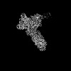
- Structure visualization
| Structure viewer | Molecule: MolmilJmol/JSmol |
|---|
- Downloads & links
-Download
| PDBx/mmCIF format | 8hbd.cif.gz | 245.2 KB | Display | PDBx/mmCIF format |
|---|---|---|---|---|
| PDB format | pdb8hbd.ent.gz | 183.7 KB | Display | PDB format |
| PDBx/mmJSON format | 8hbd.json.gz | Tree view | PDBx/mmJSON format | |
| Others |  Other downloads Other downloads |
-Validation report
| Arichive directory | https://data.pdbj.org/pub/pdb/validation_reports/hb/8hbdftp://data.pdbj.org/pub/pdb/validation_reports/hb/8hbd | HTTPS FTP |
|---|
-Related structure data
| Related structure data | 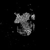 34619MC 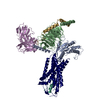 8hcqC 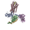 8hcxC M: map data used to model this data C: citing same article ( |
|---|---|
| Similar structure data |
-Links
 PDBj
PDBj


- Assembly
| Deposited unit | 
|
|---|---|
| 1 |
|
-Components
-Guanine nucleotide-binding protein ... , 3 types, 3 molecules ABG
| #1: Protein | Mass: 40445.059 Da / Num. of mol.: 1 Source method: isolated from a genetically manipulated source Source: (gene. exp.) Homo sapiens (human) / Gene: GNAI1 / Production host:   Spodoptera frugiperda (fall armyworm) / References: UniProt: P63096 Spodoptera frugiperda (fall armyworm) / References: UniProt: P63096 |
|---|---|
| #2: Protein | Mass: 41055.867 Da / Num. of mol.: 1 Source method: isolated from a genetically manipulated source Source: (gene. exp.) Homo sapiens (human) / Gene: GNB1 / Production host: Spodoptera frugiperda (fall armyworm) / References: UniProt: P62873 |
| #3: Protein | Mass: 7861.143 Da / Num. of mol.: 1 Source method: isolated from a genetically manipulated source Source: (gene. exp.) Homo sapiens (human) / Gene: GNG2 / Production host: Spodoptera frugiperda (fall armyworm) / References: UniProt: P59768 |
-Antibody / Protein/peptide / Protein , 3 types, 3 molecules HLR
| #4: Antibody | Mass: 30363.043 Da / Num. of mol.: 1 Source method: isolated from a genetically manipulated source Source: (gene. exp.) Spodoptera frugiperda (fall armyworm) |
|---|---|
| #5: Protein/peptide | Mass: 1836.970 Da / Num. of mol.: 1 Source method: isolated from a genetically manipulated source Source: (gene. exp.) Homo sapiens (human) / Production host: Spodoptera frugiperda (fall armyworm) |
| #6: Protein | Mass: 67882.320 Da / Num. of mol.: 1 Source method: isolated from a genetically manipulated source Source: (gene. exp.) Homo sapiens (human) / Gene: EDNRB, ETRB / Production host: Spodoptera frugiperda (fall armyworm)References: UniProt: P24530, Oplophorus-luciferin 2-monooxygenase |
-Details
| Has protein modification | Y |
|---|
-Experimental details
-Experiment
| Experiment | Method: ELECTRON MICROSCOPY |
|---|---|
| EM experiment | Aggregation state: PARTICLE / 3D reconstruction method: single particle reconstruction |
- Sample preparation
| Component | Name: IRL1620-bound ETBR-Gi complex / Type: COMPLEX / Entity ID: all / Source: MULTIPLE SOURCES | ||||||||||||
|---|---|---|---|---|---|---|---|---|---|---|---|---|---|
| Source (natural) |
| ||||||||||||
| Source (recombinant) | Organism: Spodoptera frugiperda (fall armyworm) | ||||||||||||
| Buffer solution | pH: 7.04 | ||||||||||||
| Specimen | Embedding applied: NO / Shadowing applied: NO / Staining applied: NO / Vitrification applied: YES | ||||||||||||
| Vitrification | Cryogen name: ETHANE |
- Electron microscopy imaging
| Experimental equipment |  Model: Titan Krios / Image courtesy: FEI Company |
|---|---|
| Microscopy | Model: FEI TITAN KRIOS |
| Electron gun | Electron source:  FIELD EMISSION GUN / Accelerating voltage: 300 kV / Illumination mode: FLOOD BEAM FIELD EMISSION GUN / Accelerating voltage: 300 kV / Illumination mode: FLOOD BEAM |
| Electron lens | Mode: BRIGHT FIELD / Nominal defocus max: 18000 nm / Nominal defocus min: 800 nm |
| Image recording | Electron dose: 50 e/Å2 / Film or detector model: GATAN K3 (6k x 4k) |
- Processing
| Software | Name: PHENIX / Version: 1.20.1_4487: / Classification: refinement | ||||||||||||||||||||||||
|---|---|---|---|---|---|---|---|---|---|---|---|---|---|---|---|---|---|---|---|---|---|---|---|---|---|
| CTF correction | Type: PHASE FLIPPING AND AMPLITUDE CORRECTION | ||||||||||||||||||||||||
| 3D reconstruction | Resolution: 2.99 Å / Resolution method: FSC 0.143 CUT-OFF / Num. of particles: 191493 / Symmetry type: POINT | ||||||||||||||||||||||||
| Refine LS restraints |
|
