[English] 日本語
Yorodumi Papers
- Database of articles cited by EMDB/PDB/SASBDB data -

+
Search query

Keywords
Structure methods
Author
Journal
IF

-
Structure paper

TitleStructure deformation and curvature sensing of PIEZO1 in lipid membranes.
Journal, issue, pagesNature, Vol. 604, Issue 7905, Page 377-383, Year 2022
Publish dateApr 6, 2022
AuthorsXuzhong Yang / Chao Lin / Xudong Chen / Shouqin Li / Xueming Li / Bailong Xiao /
PubMed AbstractPIEZO channels respond to piconewton-scale forces to mediate critical physiological and pathophysiological processes. Detergent-solubilized PIEZO channels form bowl-shaped trimers comprising a ...PIEZO channels respond to piconewton-scale forces to mediate critical physiological and pathophysiological processes. Detergent-solubilized PIEZO channels form bowl-shaped trimers comprising a central ion-conducting pore with an extracellular cap and three curved and non-planar blades with intracellular beams, which may undergo force-induced deformation within lipid membranes. However, the structures and mechanisms underlying the gating dynamics of PIEZO channels in lipid membranes remain unresolved. Here we determine the curved and flattened structures of PIEZO1 reconstituted in liposome vesicles, directly visualizing the substantial deformability of the PIEZO1-lipid bilayer system and an in-plane areal expansion of approximately 300 nm in the flattened structure. The curved structure of PIEZO1 resembles the structure determined from detergent micelles, but has numerous bound phospholipids. By contrast, the flattened structure exhibits membrane tension-induced flattening of the blade, bending of the beam and detaching and rotating of the cap, which could collectively lead to gating of the ion-conducting pathway. On the basis of the measured in-plane membrane area expansion and stiffness constant of PIEZO1 (ref. ), we calculate a half maximal activation tension of about 1.9 pN nm, matching experimentally measured values. Thus, our studies provide a fundamental understanding of how the notable deformability and structural rearrangement of PIEZO1 achieve exquisite mechanosensitivity and unique curvature-based gating in lipid membranes.
External linksNature / PubMed:35388220
MethodsEM (single particle)
Resolution3.46 - 6.81 Å
Structure data

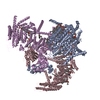
EMDB-32592, PDB-7wlt:
the Curved Structure of mPIEZO1 in Lipid Bilayer
Method: EM (single particle) / Resolution: 3.46 Å

EMDB-32593: the Curved Structure of mPIEZO1 in Lipid Bilayer
PDB-7wlu: The Flattened Structure of mPIEZO1 in Lipid Bilayer
Method: EM (single particle) / Resolution: 6.81 Å

Chemicals

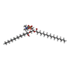
ChemComp-PLX:
(9R,11S)-9-({[(1S)-1-HYDROXYHEXADECYL]OXY}METHYL)-2,2-DIMETHYL-5,7,10-TRIOXA-2LAMBDA~5~-AZA-6LAMBDA~5~-PHOSPHAOCTACOSANE-6,6,11-TRIOL / phospholipid*YM

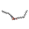
ChemComp-PEE:
1,2-dioleoyl-sn-glycero-3-phosphoethanolamine / DOPE, phospholipid*YM

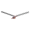
ChemComp-P5S:
O-[(R)-{[(2R)-2,3-bis(octadecanoyloxy)propyl]oxy}(hydroxy)phosphoryl]-L-serine

Source
  • mus musculus (house mouse)
KeywordsMEMBRANE PROTEIN / Force / Flexible / Complex

+
About Yorodumi Papers

-
News

-
Feb 9, 2022. New format data for meta-information of EMDB entries

New format data for meta-information of EMDB entries

  • Version 3 of the EMDB header file is now the official format.
  • The previous official version 1.9 will be removed from the archive.

Related info.:EMDB header

External links:wwPDB to switch to version 3 of the EMDB data model

-
Aug 12, 2020. Covid-19 info

Covid-19 info

URL: https://pdbj.org/emnavi/covid19.php

New page: Covid-19 featured information page in EM Navigator.

Related info.:Covid-19 info / Mar 5, 2020. Novel coronavirus structure data

+
Mar 5, 2020. Novel coronavirus structure data

Novel coronavirus structure data

Related info.:Yorodumi Speices / Aug 12, 2020. Covid-19 info

External links:COVID-19 featured content - PDBj / Molecule of the Month (242):Coronavirus Proteases

+
Jan 31, 2019. EMDB accession codes are about to change! (news from PDBe EMDB page)

EMDB accession codes are about to change! (news from PDBe EMDB page)

  • The allocation of 4 digits for EMDB accession codes will soon come to an end. Whilst these codes will remain in use, new EMDB accession codes will include an additional digit and will expand incrementally as the available range of codes is exhausted. The current 4-digit format prefixed with “EMD-” (i.e. EMD-XXXX) will advance to a 5-digit format (i.e. EMD-XXXXX), and so on. It is currently estimated that the 4-digit codes will be depleted around Spring 2019, at which point the 5-digit format will come into force.
  • The EM Navigator/Yorodumi systems omit the EMD- prefix.

Related info.:Q: What is EMD? / ID/Accession-code notation in Yorodumi/EM Navigator

External links:EMDB Accession Codes are Changing Soon! / Contact to PDBj

+
Jul 12, 2017. Major update of PDB

Major update of PDB

  • wwPDB released updated PDB data conforming to the new PDBx/mmCIF dictionary.
  • This is a major update changing the version number from 4 to 5, and with Remediation, in which all the entries are updated.
  • In this update, many items about electron microscopy experimental information are reorganized (e.g. em_software).
  • Now, EM Navigator and Yorodumi are based on the updated data.

External links:wwPDB Remediation / Enriched Model Files Conforming to OneDep Data Standards Now Available in the PDB FTP Archive

-
Yorodumi Papers

Database of articles cited by EMDB/PDB/SASBDB data

  • Database of articles cited by EMDB, PDB, and SASBDB entries
  • Using PubMed data

Related info.:EMDB / PDB / SASBDB / Yorodumi / EMN Papers / Changes in new EM Navigator and Yorodumi

Read more