+ Open data
- Basic information
| Entry | Database: PDB / ID: 7wlu | ||||||||||||||||||
|---|---|---|---|---|---|---|---|---|---|---|---|---|---|---|---|---|---|---|---|
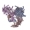
| Title | The Flattened Structure of mPIEZO1 in Lipid Bilayer | ||||||||||||||||||
 Components Components | Piezo-type mechanosensitive ion channel component 1 | ||||||||||||||||||
 Keywords Keywords | MEMBRANE PROTEIN / Force / Flexible / Complex | ||||||||||||||||||
| Function / homology |  Function and homology information Function and homology informationmechanosensitive monoatomic cation channel activity / cuticular plate / positive regulation of cell-cell adhesion mediated by integrin / positive regulation of integrin activation / detection of mechanical stimulus / mechanosensitive monoatomic ion channel activity / stereocilium / positive regulation of myotube differentiation / lamellipodium membrane / monoatomic cation transport ...mechanosensitive monoatomic cation channel activity / cuticular plate / positive regulation of cell-cell adhesion mediated by integrin / positive regulation of integrin activation / detection of mechanical stimulus / mechanosensitive monoatomic ion channel activity / stereocilium / positive regulation of myotube differentiation / lamellipodium membrane / monoatomic cation transport / monoatomic cation channel activity / endoplasmic reticulum-Golgi intermediate compartment membrane / regulation of membrane potential / endoplasmic reticulum membrane / endoplasmic reticulum / identical protein binding / plasma membrane Similarity search - Function | ||||||||||||||||||
| Biological species |  | ||||||||||||||||||
| Method | ELECTRON MICROSCOPY / single particle reconstruction / cryo EM / Resolution: 6.81 Å | ||||||||||||||||||
 Authors Authors | Yang, X. / Lin, C. / Chen, X. / Li, S. / Li, X. / Xiao, B. | ||||||||||||||||||
| Funding support |  China, 5items China, 5items
| ||||||||||||||||||
 Citation Citation |  Journal: Nature / Year: 2022 Journal: Nature / Year: 2022Title: Structure deformation and curvature sensing of PIEZO1 in lipid membranes. Authors: Xuzhong Yang / Chao Lin / Xudong Chen / Shouqin Li / Xueming Li / Bailong Xiao /  Abstract: PIEZO channels respond to piconewton-scale forces to mediate critical physiological and pathophysiological processes. Detergent-solubilized PIEZO channels form bowl-shaped trimers comprising a ...PIEZO channels respond to piconewton-scale forces to mediate critical physiological and pathophysiological processes. Detergent-solubilized PIEZO channels form bowl-shaped trimers comprising a central ion-conducting pore with an extracellular cap and three curved and non-planar blades with intracellular beams, which may undergo force-induced deformation within lipid membranes. However, the structures and mechanisms underlying the gating dynamics of PIEZO channels in lipid membranes remain unresolved. Here we determine the curved and flattened structures of PIEZO1 reconstituted in liposome vesicles, directly visualizing the substantial deformability of the PIEZO1-lipid bilayer system and an in-plane areal expansion of approximately 300 nm in the flattened structure. The curved structure of PIEZO1 resembles the structure determined from detergent micelles, but has numerous bound phospholipids. By contrast, the flattened structure exhibits membrane tension-induced flattening of the blade, bending of the beam and detaching and rotating of the cap, which could collectively lead to gating of the ion-conducting pathway. On the basis of the measured in-plane membrane area expansion and stiffness constant of PIEZO1 (ref. ), we calculate a half maximal activation tension of about 1.9 pN nm, matching experimentally measured values. Thus, our studies provide a fundamental understanding of how the notable deformability and structural rearrangement of PIEZO1 achieve exquisite mechanosensitivity and unique curvature-based gating in lipid membranes. | ||||||||||||||||||
| History |
|
- Structure visualization
| Structure viewer | Molecule:  Molmil Molmil Jmol/JSmol Jmol/JSmol |
|---|
- Downloads & links
- Download
| PDBx/mmCIF format |  7wlu.cif.gz 7wlu.cif.gz | 825.4 KB | Display |  PDBx/mmCIF format PDBx/mmCIF format |
|---|---|---|---|---|
| PDB format |  pdb7wlu.ent.gz pdb7wlu.ent.gz | 639.1 KB | Display |  PDB format PDB format |
| PDBx/mmJSON format |  7wlu.json.gz 7wlu.json.gz | Tree view |  PDBx/mmJSON format PDBx/mmJSON format | |
| Others |  Other downloads Other downloads |
-Validation report
| Arichive directory |  https://data.pdbj.org/pub/pdb/validation_reports/wl/7wlu https://data.pdbj.org/pub/pdb/validation_reports/wl/7wlu ftp://data.pdbj.org/pub/pdb/validation_reports/wl/7wlu ftp://data.pdbj.org/pub/pdb/validation_reports/wl/7wlu | HTTPS FTP |
|---|
-Related structure data
| Related structure data |  32593MC  7wltC M: map data used to model this data C: citing same article ( |
|---|---|
| Similar structure data | Similarity search - Function & homology  F&H Search F&H Search |
- Links
Links
- Assembly
| Deposited unit | 
|
|---|---|
| 1 |
|
- Components
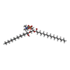
| #1: Protein | Mass: 292320.656 Da / Num. of mol.: 3 Source method: isolated from a genetically manipulated source Source: (gene. exp.)   Homo sapiens (human) / References: UniProt: E2JF22 Homo sapiens (human) / References: UniProt: E2JF22#2: Chemical | Has ligand of interest | Y | |
|---|
-Experimental details
-Experiment
| Experiment | Method: ELECTRON MICROSCOPY |
|---|---|
| EM experiment | Aggregation state: PARTICLE / 3D reconstruction method: single particle reconstruction |
- Sample preparation
| Component | Name: The Flattened Structure of mPIEZO1 in Lipid Bilayer / Type: COMPLEX / Entity ID: #1 / Source: RECOMBINANT |
|---|---|
| Source (natural) | Organism:  |
| Source (recombinant) | Organism:  Homo sapiens (human) Homo sapiens (human) |
| Buffer solution | pH: 7.2 |
| Specimen | Embedding applied: NO / Shadowing applied: NO / Staining applied: NO / Vitrification applied: YES |
| Vitrification | Cryogen name: ETHANE |
- Electron microscopy imaging
| Experimental equipment |  Model: Titan Krios / Image courtesy: FEI Company |
|---|---|
| Microscopy | Model: FEI TITAN KRIOS |
| Electron gun | Electron source:  FIELD EMISSION GUN / Accelerating voltage: 300 kV / Illumination mode: FLOOD BEAM FIELD EMISSION GUN / Accelerating voltage: 300 kV / Illumination mode: FLOOD BEAM |
| Electron lens | Mode: BRIGHT FIELD / Nominal defocus max: 2400 nm / Nominal defocus min: 1500 nm |
| Image recording | Electron dose: 50 e/Å2 / Film or detector model: GATAN K3 (6k x 4k) |
- Processing
| CTF correction | Type: NONE |
|---|---|
| 3D reconstruction | Resolution: 6.81 Å / Resolution method: FSC 0.143 CUT-OFF / Num. of particles: 35012 / Symmetry type: POINT |
 Movie
Movie Controller


 PDBj
PDBj


