 Movie
Movie Controller


+ Open data

- Basic information
| Entry |  | ||||||||||||||||||
|---|---|---|---|---|---|---|---|---|---|---|---|---|---|---|---|---|---|---|---|
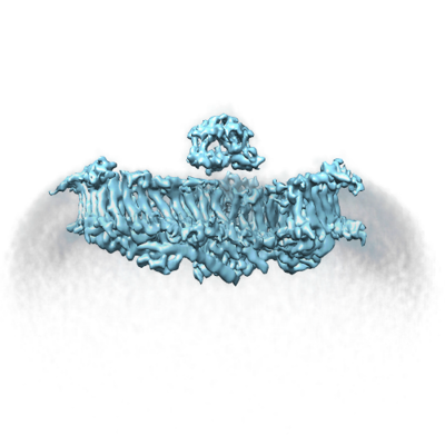
| Title | the Curved Structure of mPIEZO1 in Lipid Bilayer | ||||||||||||||||||
 Map data Map data | |||||||||||||||||||
 Sample Sample |
| ||||||||||||||||||
 Keywords Keywords | Force / Flexible / Complex / MEMBRANE PROTEIN | ||||||||||||||||||
| Function / homology |  Function and homology information Function and homology informationmechanosensitive monoatomic cation channel activity / cuticular plate / positive regulation of integrin activation / detection of mechanical stimulus / positive regulation of cell-cell adhesion mediated by integrin / mechanosensitive monoatomic ion channel activity / stereocilium / positive regulation of myotube differentiation / lamellipodium membrane / monoatomic cation transport ...mechanosensitive monoatomic cation channel activity / cuticular plate / positive regulation of integrin activation / detection of mechanical stimulus / positive regulation of cell-cell adhesion mediated by integrin / mechanosensitive monoatomic ion channel activity / stereocilium / positive regulation of myotube differentiation / lamellipodium membrane / monoatomic cation transport / monoatomic cation channel activity / endoplasmic reticulum-Golgi intermediate compartment membrane / cellular response to mechanical stimulus / regulation of membrane potential / endoplasmic reticulum membrane / endoplasmic reticulum / identical protein binding / plasma membrane Similarity search - Function | ||||||||||||||||||
| Biological species |  | ||||||||||||||||||
| Method | single particle reconstruction / cryo EM / Resolution: 6.81 Å | ||||||||||||||||||
 Authors Authors | Yang X / Lin C / Chen X / Li S / Li X / Xiao B | ||||||||||||||||||
| Funding support |  China, 5 items China, 5 items
| ||||||||||||||||||
 Citation Citation | Journal: Nature / Year: 2022 Title: Structure deformation and curvature sensing of PIEZO1 in lipid membranes. Authors: Xuzhong Yang / Chao Lin / Xudong Chen / Shouqin Li / Xueming Li / Bailong Xiao / Abstract: PIEZO channels respond to piconewton-scale forces to mediate critical physiological and pathophysiological processes. Detergent-solubilized PIEZO channels form bowl-shaped trimers comprising a ...PIEZO channels respond to piconewton-scale forces to mediate critical physiological and pathophysiological processes. Detergent-solubilized PIEZO channels form bowl-shaped trimers comprising a central ion-conducting pore with an extracellular cap and three curved and non-planar blades with intracellular beams, which may undergo force-induced deformation within lipid membranes. However, the structures and mechanisms underlying the gating dynamics of PIEZO channels in lipid membranes remain unresolved. Here we determine the curved and flattened structures of PIEZO1 reconstituted in liposome vesicles, directly visualizing the substantial deformability of the PIEZO1-lipid bilayer system and an in-plane areal expansion of approximately 300 nm in the flattened structure. The curved structure of PIEZO1 resembles the structure determined from detergent micelles, but has numerous bound phospholipids. By contrast, the flattened structure exhibits membrane tension-induced flattening of the blade, bending of the beam and detaching and rotating of the cap, which could collectively lead to gating of the ion-conducting pathway. On the basis of the measured in-plane membrane area expansion and stiffness constant of PIEZO1 (ref. ), we calculate a half maximal activation tension of about 1.9 pN nm, matching experimentally measured values. Thus, our studies provide a fundamental understanding of how the notable deformability and structural rearrangement of PIEZO1 achieve exquisite mechanosensitivity and unique curvature-based gating in lipid membranes. | ||||||||||||||||||
| History |
|
- Structure visualization
| Supplemental images |
|---|
- Downloads & links
-EMDB archive
| Map data | emd_32593.map.gz | 20.4 MB | EMDB map data format | |
|---|---|---|---|---|
| Header (meta data) | emd-32593-v30.xmlemd-32593.xml | 12.8 KB 12.8 KB | Display Display | EMDB header |
| Images |  emd_32593.png emd_32593.png | 92.4 KB | ||
| Filedesc metadata | emd-32593.cif.gz | 6.7 KB | ||
| Archive directory |  http://ftp.pdbj.org/pub/emdb/structures/EMD-32593ftp://ftp.pdbj.org/pub/emdb/structures/EMD-32593 http://ftp.pdbj.org/pub/emdb/structures/EMD-32593ftp://ftp.pdbj.org/pub/emdb/structures/EMD-32593 | HTTPS FTP |
-Related structure data
| Related structure data |  7wluMC  7wltC M: atomic model generated by this map C: citing same article ( |
|---|---|
| Similar structure data |

-Links
| EMDB pages | EMDB (EBI/PDBe) / EMDataResource |
|---|---|
| Related items in Molecule of the Month |

-Map
| File | Download / File: emd_32593.map.gz / Format: CCP4 / Size: 22.2 MB / Type: IMAGE STORED AS FLOATING POINT NUMBER (4 BYTES) | ||||||||||||||||||||||||||||||||||||
|---|---|---|---|---|---|---|---|---|---|---|---|---|---|---|---|---|---|---|---|---|---|---|---|---|---|---|---|---|---|---|---|---|---|---|---|---|---|
| Projections & slices | Image control
Images are generated by Spider. | ||||||||||||||||||||||||||||||||||||
| Voxel size | X=Y=Z: 2.196 Å | ||||||||||||||||||||||||||||||||||||
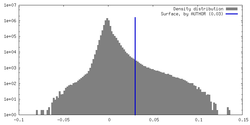
| Density |
| ||||||||||||||||||||||||||||||||||||
| Symmetry | Space group: 1 | ||||||||||||||||||||||||||||||||||||
| Details | EMDB XML:
|
 Z (Sec.)
 Y (Row.)
 X (Col.)


-Supplemental data
- Sample components
-Entire : The Flattened Structure of mPIEZO1 in Lipid Bilayer
| Entire | Name: The Flattened Structure of mPIEZO1 in Lipid Bilayer |
|---|---|
| Components |
|
-Supramolecule #1: The Flattened Structure of mPIEZO1 in Lipid Bilayer
| Supramolecule | Name: The Flattened Structure of mPIEZO1 in Lipid Bilayer / type: complex / ID: 1 / Parent: 0 / Macromolecule list: #1 |
|---|---|
| Source (natural) | Organism: |
-Macromolecule #1: Piezo-type mechanosensitive ion channel component 1
| Macromolecule | Name: Piezo-type mechanosensitive ion channel component 1 / type: protein_or_peptide / ID: 1 / Number of copies: 3 / Enantiomer: LEVO |
|---|---|
| Source (natural) | Organism: |
| Molecular weight | Theoretical: 292.320656 KDa |
| Recombinant expression | Organism:  Homo sapiens (human) Homo sapiens (human) |
| Sequence | String: MEPHVLGAGL YWLLLPCTLL AASLLRFNAL SLVYLLFLLL LPWLPGPSRH SIPGHTGRLL RALLCLSLLF LVAHLAFQIC LHTVPHLDQ FLGQNGSLWV KVSQHIGVTR LDLKDIFNTT RLVAPDLGVL LASSLCLGLC GRLTRKAGQS RRTQELQDDD D DDDDDDED ...String: MEPHVLGAGL YWLLLPCTLL AASLLRFNAL SLVYLLFLLL LPWLPGPSRH SIPGHTGRLL RALLCLSLLF LVAHLAFQIC LHTVPHLDQ FLGQNGSLWV KVSQHIGVTR LDLKDIFNTT RLVAPDLGVL LASSLCLGLC GRLTRKAGQS RRTQELQDDD D DDDDDDED IDAAPAVGLK GAPALATKRR LWLASRFRVT AHWLLMTSGR TLVIVLLALA GIAHPSAFSS IYLVVFLAIC TW WSCHFPL SPLGFNTLCV MVSCFGAGHL ICLYCYQTPF IQDMLPPGNI WARLFGLKNF VDLPNYSSPN ALVLNTKHAW PIY VSPGIL LLLYYTATSL LKLHKSCPSE LRKETPREDE EHELELDHLE PEPQARDATQ GEMPMTTEPD LDNCTVHVLT SQSP VRQRP VRPRLAELKE MSPLHGLGHL IMDQSYVCAL IAMMVWSIMY HSWLTFVLLL WACLIWTVRS RHQLAMLCSP CILLY GLTL CCLRYVWAME LPELPTTLGP VSLHQLGLEH TRYPCLDLGA MLLYLLTFWL LLRQFVKEKL LKKQKVPAAL LEVTVA DTE PTQTQTLLRS LGELVTGIYV KYWIYVCAGM FIVVSFAGRL VVYKIVYMFL FLLCLTLFQV YYTLWRKLLR VFWWLVV AY TMLVLIAVYT FQFQDFPTYW RNLTGFTDEQ LGDLGLEQFS VSELFSSILI PGFFLLACIL QLHYFHRPFM QLTDLEHV P PPGTRHPRWA HRQDAVSEAP LLEHQEEEEV FREDGQSMDG PHQATQVPEG TASKWGLVAD RLLDLAASFS AVLTRIQVF VRRLLELHVF KLVALYTVWV ALKEVSVMNL LLVVLWAFAL PYPRFRPMAS CLSTVWTCII IVCKMLYQLK IVNPHEYSSN CTEPFPNNT NLQPLEINQS LLYRGPVDPA NWFGVRKGYP NLGYIQNHLQ ILLLLVFEAV VYRRQEHYRR QHQQAPLPAQ A VCADGTRQ RLDQDLLSCL KYFINFFFYK FGLEICFLMA VNVIGQRMNF MVILHGCWLV AILTRRRREA IARLWPNYCL FL TLFLLYQ YLLCLGMPPA LCIDYPWRWS KAIPMNSALI KWLYLPDFFR APNSTNLISD FLLLLCASQQ WQVFSAERTE EWQ RMAGIN TDHLEPLRGE PNPIPNFIHC RSYLDMLKVA VFRYLFWLVL VVVFVAGATR ISIFGLGYLL ACFYLLLFGT TLLQ KDTRA QLVLWDCLIL YNVTVIISKN MLSLLSCVFV EQMQSNFCWV IQLFSLVCTV KGYYDPKEMM TRDRDCLLPV EEAGI IWDS ICFFFLLLQR RIFLSHYFLH VSADLKATAL QASRGFALYN AANLKSINFH RQIEEKSLAQ LKRQMKRIRA KQEKYR QSQ ASRGQLQSKD PQDPSQEPGP DSPGGSSPPR RQWWRPWLDH ATVIHSGDYF LFESDSEEEE EALPEDPRPA AQSAFQM AY QAWVTNAQTV LRQRRERARQ ERAEQLASGG DLNPDVEPVD VPEDEMAGRS HMMQRVLSTM QFLWVLGQAT VDGLTRWL R AFTKHHRTMS DVLCAERYLL TQELLRVGEV RRGVLDQLYV GEDEATLSGP VETRDGPSTA SSGLGAEEPL SSMTDDTSS PLSTGYNTRS GSEEIVTDAG DLQAGTSLHG SQELLANART RMRTASELLL DRRLHIPELE EAERFEAQQG RTLRLLRAGY QCVAAHSEL LCYFIIILNH MVTASAASLV LPVLVFLWAM LTIPRPSKRF WMTAIVFTEV MVVTKYLFQF GFFPWNSYVV L RRYENKPY FPPRILGLEK TDSYIKYDLV QLMALFFHRS QLLCYGLWDH EEDRYPKDHC RSSVKDREAK EEPEAKLESQ SE TGTGHPK EPVLAGTPRD HIQGKGSIRS KDVIQDPPED LKPRHTRHIS IRFRRRKETP GPKGTAVMET EHEEGEGKET TER KRPRHT QEKSKFRERM KAAGRRLQSF CVSLAQSFYQ PLQRFFHDIL HTKYRAATDV YALMFLADIV DIIIIIFGFW AFGK HSAAT DIASSLSDDQ VPQAFLFMLL VQFGTMVIDR ALYLRKTVLG KLAFQVVLVV AIHIWMFFIL PAVTERMFSQ NAVAQ LWYF VKCIYFALSA YQIRCGYPTR ILGNFLTKKY NHLNLFLFQG FRLVPFLVEL RAVMDWVWTD TTLSLSNWMC VEDIYA NIF IIKCSRETEK KYPQPKGQKK KKIVKYGMGG LIILFLIAII WFPLLFMSLI RSVVGVVNQP IDVTVTLKLG GYEPLFT MS AQQPSIVPFT PQAYEELSQQ FDPYPLAMQF ISQYSPEDIV TAQIEGSSGA LWRISPPSRA QMKQELYNGT ADITLRFT W NFQRDLAKGG TVEYTNEKHT LELAPNSTAR RQLAQLLEGR PDQSVVIPHL FPKYIRAPNG PEANPVKQLQ PDEEEDYLG VRIQLRREQV GTGASGEQAG TKASDFLEWW VIELQDCKAD CNLLPMVIFS DKVSPPSLGF LAGYGIVGLY VSIVLVVGKF VRGFFSEIS HSIMFEELPC VDRILKLCQD IFLVRETREL ELEEELYAKL IFLYRSPETM IKWTRERE UniProtKB: Piezo-type mechanosensitive ion channel component 1 |
-Macromolecule #2: (9R,11S)-9-({[(1S)-1-HYDROXYHEXADECYL]OXY}METHYL)-2,2-DIMETHYL-5,...
| Macromolecule | Name: (9R,11S)-9-({[(1S)-1-HYDROXYHEXADECYL]OXY}METHYL)-2,2-DIMETHYL-5,7,10-TRIOXA-2LAMBDA~5~-AZA-6LAMBDA~5~-PHOSPHAOCTACOSANE-6,6,11-TRIOL type: ligand / ID: 2 / Number of copies: 3 / Formula: PLX |
|---|---|
| Molecular weight | Theoretical: 767.132 Da |
| Chemical component information |  ChemComp-PLX: |
-Experimental details
-Structure determination
| Method | cryo EM |
|---|---|
 Processing Processing | single particle reconstruction |
| Aggregation state | particle |
-Sample preparation
| Buffer | pH: 7.2 |
|---|---|
| Vitrification | Cryogen name: ETHANE |
- Electron microscopy
| Microscope | FEI TITAN KRIOS |
|---|---|
| Image recording | Film or detector model: GATAN K3 (6k x 4k) / Average electron dose: 50.0 e/Å2 |
| Electron beam | Acceleration voltage: 300 kV / Electron source:  FIELD EMISSION GUN FIELD EMISSION GUN |
| Electron optics | Illumination mode: FLOOD BEAM / Imaging mode: BRIGHT FIELD / Nominal defocus max: 2.4 µm / Nominal defocus min: 1.5 µm |
| Experimental equipment |  Model: Titan Krios / Image courtesy: FEI Company |
-Image processing
| Startup model | Type of model: EMDB MAP EMDB ID: |
|---|---|
| Final reconstruction | Resolution.type: BY AUTHOR / Resolution: 6.81 Å / Resolution method: FSC 0.143 CUT-OFF / Number images used: 35012 |
| Initial angle assignment | Type: MAXIMUM LIKELIHOOD |
| Final angle assignment | Type: MAXIMUM LIKELIHOOD |

