ムービー
ムービー コントローラー
 構造ビューア
 万見文献について


+検索条件
-Structure paper
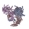
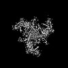
| タイトル | Structure deformation and curvature sensing of PIEZO1 in lipid membranes. |
|---|---|
| ジャーナル・号・ページ | Nature, Vol. 604, Issue 7905, Page 377-383, Year 2022 |
| 掲載日 | 2022年4月6日 |
 著者 著者 | Xuzhong Yang / Chao Lin / Xudong Chen / Shouqin Li / Xueming Li / Bailong Xiao /  |
| PubMed 要旨 | PIEZO channels respond to piconewton-scale forces to mediate critical physiological and pathophysiological processes. Detergent-solubilized PIEZO channels form bowl-shaped trimers comprising a ...PIEZO channels respond to piconewton-scale forces to mediate critical physiological and pathophysiological processes. Detergent-solubilized PIEZO channels form bowl-shaped trimers comprising a central ion-conducting pore with an extracellular cap and three curved and non-planar blades with intracellular beams, which may undergo force-induced deformation within lipid membranes. However, the structures and mechanisms underlying the gating dynamics of PIEZO channels in lipid membranes remain unresolved. Here we determine the curved and flattened structures of PIEZO1 reconstituted in liposome vesicles, directly visualizing the substantial deformability of the PIEZO1-lipid bilayer system and an in-plane areal expansion of approximately 300 nm in the flattened structure. The curved structure of PIEZO1 resembles the structure determined from detergent micelles, but has numerous bound phospholipids. By contrast, the flattened structure exhibits membrane tension-induced flattening of the blade, bending of the beam and detaching and rotating of the cap, which could collectively lead to gating of the ion-conducting pathway. On the basis of the measured in-plane membrane area expansion and stiffness constant of PIEZO1 (ref. ), we calculate a half maximal activation tension of about 1.9 pN nm, matching experimentally measured values. Thus, our studies provide a fundamental understanding of how the notable deformability and structural rearrangement of PIEZO1 achieve exquisite mechanosensitivity and unique curvature-based gating in lipid membranes. |
 リンク リンク | Nature / PubMed:35388220 |
| 手法 | EM (単粒子) |
| 解像度 | 3.46 - 6.81 Å |
| 構造データ | EMDB-32592, PDB-7wlt: EMDB-32593: the Curved Structure of mPIEZO1 in Lipid Bilayer |
| 化合物 | 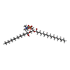 ChemComp-PLX:  ChemComp-PEE:  ChemComp-P5S: |
| 由来 |
|
 キーワード キーワード | MEMBRANE PROTEIN / Force / Flexible / Complex |