Movie
Movie Controller
 Structure viewers
 About Yorodumi Papers


+Search query
-Structure paper
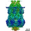
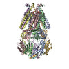
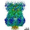
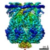
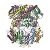
| Title | Visualization of the mechanosensitive ion channel MscS under membrane tension. |
|---|---|
| Journal, issue, pages | Nature, Vol. 590, Issue 7846, Page 509-514, Year 2021 |
| Publish date | Feb 10, 2021 |
 Authors Authors | Yixiao Zhang / Csaba Daday / Ruo-Xu Gu / Charles D Cox / Boris Martinac / Bert L de Groot / Thomas Walz /    |
| PubMed Abstract | Mechanosensitive channels sense mechanical forces in cell membranes and underlie many biological sensing processes. However, how exactly they sense mechanical force remains under investigation. The ...Mechanosensitive channels sense mechanical forces in cell membranes and underlie many biological sensing processes. However, how exactly they sense mechanical force remains under investigation. The bacterial mechanosensitive channel of small conductance, MscS, is one of the most extensively studied mechanosensitive channels, but how it is regulated by membrane tension remains unclear, even though the structures are known for its open and closed states. Here we used cryo-electron microscopy to determine the structure of MscS in different membrane environments, including one that mimics a membrane under tension. We present the structures of MscS in the subconducting and desensitized states, and demonstrate that the conformation of MscS in a lipid bilayer in the open state is dynamic. Several associated lipids have distinct roles in MscS mechanosensation. Pore lipids are necessary to prevent ion conduction in the closed state. Gatekeeper lipids stabilize the closed conformation and dissociate with membrane tension, allowing the channel to open. Pocket lipids in a solvent-exposed pocket between subunits are pulled out under sustained tension, allowing the channel to transition to the subconducting state and then to the desensitized state. Our results provide a mechanistic underpinning and expand on the 'force-from-lipids' model for MscS mechanosensation. |
 External links External links | Nature / PubMed:33568813 |
| Methods | EM (single particle) |
| Resolution | 3.2 - 3.7 Å |
| Structure data | EMDB-21462, PDB-6vyk: EMDB-21463, PDB-6vyl: EMDB-21464, PDB-6vym:  EMDB-21465: 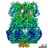 EMDB-21466: |
| Chemicals |  ChemComp-PCW: |
| Source |
|
 Keywords Keywords | MEMBRANE PROTEIN / Mechanosensitive channel / MscS / Nanodiscs / Lipid |